

ABSTRACT
Over the last 20 years, the larva of the greater waxmoth, Galleria mellonella, has rapidly increased in popularity as an in vivo mammalian replacement model organism for the study of human pathogens. Experimental readouts of response to infection are most often limited to observing the melanization cascade and quantifying larval death and, whilst transcriptomic and proteomic approaches, and methods to determine microbial load are also used, a more comprehensive toolkit of profiling infection over time could transform the applicability of this model. As an invertebrate, Galleria harbour an innate immune system comprised of both humoral components and a repertoire of innate immune cells – termed haemocytes. Although information on subtypes of haemocytes exists, there are conflicting reports on their exact number and function. Flow cytometry has previously been used to assay Galleria haemocytes, but protocols include both centrifugation and fixation – physical methods which have the potential to affect haemocyte morphology prior to analysis. Here, we present a method for live haemocyte analysis by flow cytometry, revealing that Galleria haemocytes constitute only a single resolvable population, based on relative size or internal complexity. Using fluorescent zymosan particles, we extend our method to show that up to 80% of the Galleria haemocyte population display phagocytic capability. Finally, we demonstrate that the developed assay reliably replicates in vitro data, showing that cell wall β-1,3-glucan masking by Candida albicans subverts phagocytic responses. As such, our method provides a new tool with which to rapidly assess phagocytosis and understand live infection dynamics in Galleria.
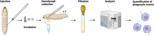
GRAPHICAL ABSTRACT
Acknowledgements
We would like to Ivan Canada Luna for his help in maintenance of the Galleria laboratory colony, and for stimulating discussions. We also thank Remy Chait (Biosciences, University of Exeter) for his provision of the E. coli strains used in this work. This work was funded by an NC3Rs Project Grant awarded to JGW and AJPB (NC/T001518/1), which supported JSC, and an NC3R Training Fellowship, awarded to JP (NC/W002388/1). AJPB was also funded by grants from the Medical Research Council UK (MR/M026663/2) and Wellcome (224323/Z/21/Z), and AP and AJPB were supported by the Medical Research Council Centre for Medical Mycology (MR/N006364/2).
Disclosure statement
No potential conflict of interest was reported by the author(s).
Contributor roles
JSC – conceptualization, methodology, investigation, formal analysis, project administration and writing (review & editing); JCP – conceptualization and writing (editing); AB – methodology, formal analysis, writing (review & editing); AP: methodology, resources, writing (review & editing); RY – methodology, formal analysis, writing (review & editing); AJPB – conceptualization, methodology, resources, writing (review & editing), supervision, funding acquisition; JGW – conceptualization, methodology, project administration, writing (review & editing), supervision, funding acquisition.
Data Availability statement
Raw data were generated at the Exeter Centre for Cytomics, UK. Derived data supporting the findings of this study are available from the corresponding author JGW/JSC on request.
Supplementary Material
Supplemental data for this article can be accessed online at https://doi.org/10.1080/21505594.2024.2313413