

ABSTRACT
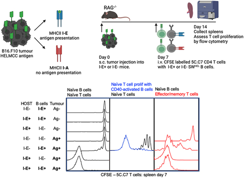
There has been growing interest in the role of B cells in antitumour immunity and potential use in adoptive cellular therapies. To date, the success of such therapies is limited. The intrinsic capacity of B cells to specifically activate tumour-specific CD4+ T cells in vivo via TCR-dependent interactions remains poorly defined. We have developed an in vivo tumour model that utilizes MHCII I-E restriction which limits antigen presentation to tumour-specific CD4 T cells to either tumour-specific B cells or host myeloid antigen presenting cells (APCs) in lymphopenic RAG-/-mice. We have previously shown that these naive tumour-specific CD4+ T cells can successfully eradicate established tumours in this model when activated by host APCs. When naïve tumour-specific B cells are the only source of I-E+ APC, very limited proliferation of naïve CD4+ T cells is observed, whereas host I-E+ APCs are potent T cell activators. B cells pre-activated with an anti-CD40 agonistic antibody in vivo support increased T cell proliferation, although far less than host APCs. CD4+ T cells that have already differentiated to an effector/central memory phenotype proliferate more readily in response to naïve B cells, although still 100-fold less than in response to host APCs. This study demonstrates that even in a significantly lymphopenic environment, myeloid APCs are the dominant primary activators of tumour-specific T cells, in contrast to the very limited capacity of tumour-specific B cells. This suggests that future anti-tumour therapies that incorporate activated B cells should also include mechanisms that activate host APCs.
GRAPHICAL ABSTRACT
Acknowledgments
We thank C. Jolly for providing HEL-Alexa647 and scientific discussion; the staff of the Centenary Institute Flow Cytometry and Animal Facilities for technical support, and members of the T cell biology and Immune Imaging Labs for scientific discussion. This work was supported by Australian National Health and Medical Research Council grants 1001020, 1012930 and 1051843 and a New South Wales Cancer Council grant RG 13-13.
Disclosure statement
No potential conflict of interest was reported by the author(s).
Data availability statement
Raw data were generated at the Centenary Institute. Derived data supporting the findings of this study are available from the corresponding author BF on request.
Supplementary material
Supplemental data for this article can be accessed online at https://doi.org/10.1080/2162402X.2023.2290799