Figures & data
Fig. 1: Copy of the bronze cast of the seal stamp ‘(belonging) to ShemaꜤ servant of Jeroboam’ from Schumacher’s excavation in Megiddo (photo by Michael Magen, the Israel Museum, Jerusalem)
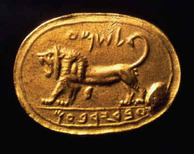
Fig. 2: The bulla, ‘(belonging) to ShemaꜤ servant of Jeroboam’; a) face; b) back (photos by Y. Goren)
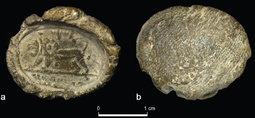
Fig. 3: a) View of the lower panel under stereomicroscope; b) the reconstructed reading (photo by Y. Goren)
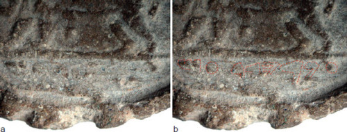
Fig. 4: RTI photo of the bulla (photo by M. Magen, the Israel Museum, Jerusalem)
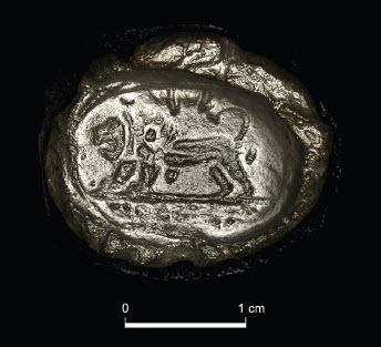
Fig. 5: Specimen from the bulla viewed under a petrographic polarising microscope (the black area marks the background); scale: 0.2 mm long; the brown silt includes limestone particles with calcite characteristic of rendzina soil; larger particles are of sand and silt containing limestone (LS), quartz (Q) and olivine (OL)
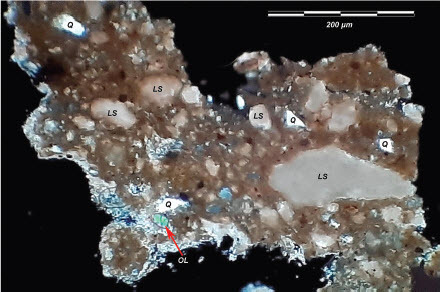
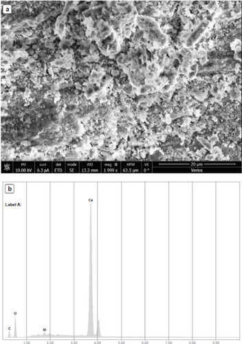
Fig. 6: Calcitic patina on the surface of the bulla’s impression examined under the SEM; a) patina formed by the accumulation of calcite crystals (scale: 0.2 mm long; b) chemical analysis of the material (a), using X-ray dispersion spectroscopy (EDS), indicating clean composition of calcite (CaCO3)