711
Views
0
CrossRef citations to date
0
Altmetric
Research Article

Onychopapilloma: a single medical center experience in Southern Taiwan

, &
Article: 2318353 | Received 20 Nov 2023, Accepted 02 Jan 2024, Published online: 28 Feb 2024

Figures & data

Figure 1. The longitudinal excision of the onychopapilloma and histological characteristics. (a) There is longitudinal leukonychia with onycholysis and distal fissuring on the right thumb. (b) Following the avulsion of the nail plate, the onychopapilloma was exposed on the nail bed, and we removed it via longitudinal excision. And the defect was closed with a non-absorbable thread. (c) The schematic diagram of the surgical procedure. The left side shows a presurgical nail lesion. The right side demonstrates the surgical outcome with suturing over the proximal nail fold and nail plate. (d) Papillomatosis and acanthosis of the nail bed, distal subungual hyperkeratosis with parakeratosis. (e) Matrix metaplasia with abundant eosinophilic cytoplasm in the upper part of nail bed epithelium.

Figure 1. The longitudinal excision of the onychopapilloma and histological characteristics. (a) There is longitudinal leukonychia with onycholysis and distal fissuring on the right thumb. (b) Following the avulsion of the nail plate, the onychopapilloma was exposed on the nail bed, and we removed it via longitudinal excision. And the defect was closed with a non-absorbable thread. (c) The schematic diagram of the surgical procedure. The left side shows a presurgical nail lesion. The right side demonstrates the surgical outcome with suturing over the proximal nail fold and nail plate. (d) Papillomatosis and acanthosis of the nail bed, distal subungual hyperkeratosis with parakeratosis. (e) Matrix metaplasia with abundant eosinophilic cytoplasm in the upper part of nail bed epithelium.

Figure 2. Clinical features of onychopapilloma. (a) The photos were taken of a 55-year-old man who presented with a five-year history of various nail abnormalities affecting both his fingernails and toenails. These abnormalities included multiple longitudinal leukonychia with distal subungual hyperkeratosis as well as focal onycholysis and splinter hemorrhage. (b) The fingernails showed multiple longitudinal leukonychia of the previous patient. (c) A solitary onychopapilloma presented wide longitudinal leukonychia with distal onycholysis and splinter hemorrhage on the ring finger. (d) A solitary onychopapilloma presented longitudinal erythronychia with distal onycholysis on the middle finger.

Figure 2. Clinical features of onychopapilloma. (a) The photos were taken of a 55-year-old man who presented with a five-year history of various nail abnormalities affecting both his fingernails and toenails. These abnormalities included multiple longitudinal leukonychia with distal subungual hyperkeratosis as well as focal onycholysis and splinter hemorrhage. (b) The fingernails showed multiple longitudinal leukonychia of the previous patient. (c) A solitary onychopapilloma presented wide longitudinal leukonychia with distal onycholysis and splinter hemorrhage on the ring finger. (d) A solitary onychopapilloma presented longitudinal erythronychia with distal onycholysis on the middle finger.

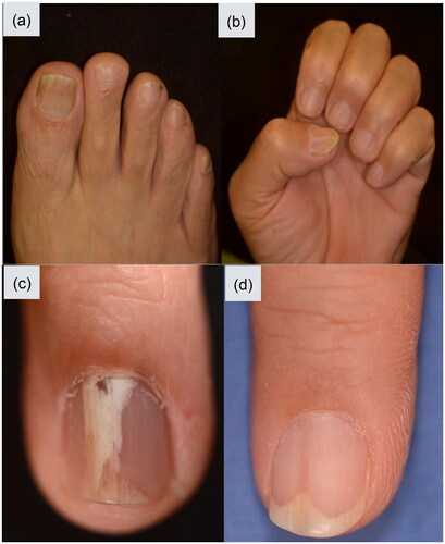
Table 1. All patients included in our study.

Table 2. Clinical presentation of onychopapilloma in our study compared with previous research.