Figures & data
Table 1. List of fungal isolates.
Figure 1. Vertical growth characteristics of the fungal isolates. Fungal isolates were inoculated at the tip of the serological pipette (25 ml, 34.5 cm) which was filled with PDA medium. The vertical growth was measured from the tip to the mycelial front propagated inside the pipette after 3 weeks of incubation at 25 °C.
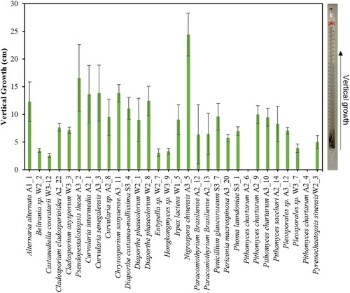
Figure 2. Growth of the fungal isolates on the minimal agar plates containing 5 g/L biological polymers, including cellulose, starch, skim milk, poly galacturonic acid and xylan. The relative growth was calculated by dividing the mycelial colony diameter grown on the biological polymers by the mycelial diameter grown on PDA.
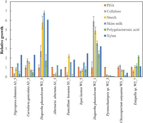
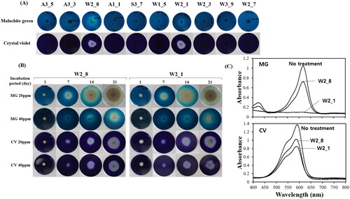
Figure 3. Degradation of malachite green (MG) and crystal violet (CV) by the fungal isolates. (A) Growth of some selected fungal isolates on PDA containing 40 ppm MG or CV. The plates were incubated 2 weeks at 25 °C. (B) Growth of Diaporthe phaseolorum W2_8 or W2_1 in different concentration of MG or CV. (C) Degradation of MG and CV by D. phaseolorum W2_8 or W2_1 in liquid culture. The mycelial cell were grown in PDA medium containing 20 ppm of MG or CV for 21 d at 25 °C. The culture supernatants, clarified by centrifugation for 5 min at 3000 rpm, were subjected to the UV-Vis spectral analysis.