Figures & data
Figure 1 A twig showing floral characters of the plant.
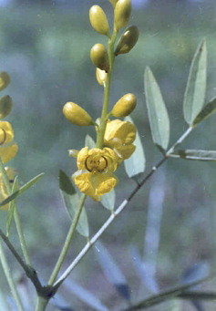
Figure 2 Macroscopic characters of seed.
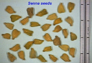
Figure 3 SEM view of whole seed.
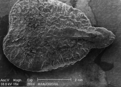
Figure 4 Surface characters of distal end showing papillae in SEM.
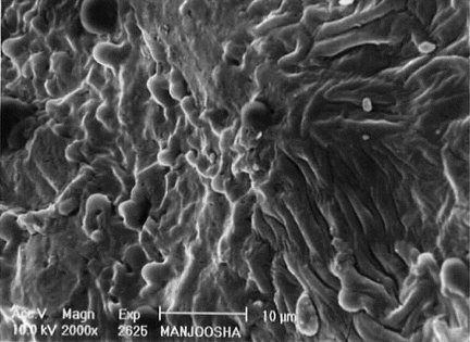
Figure 5 Surface ornamentation of seed through SEM.
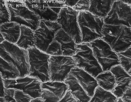
Figure 6 Inner cells of seed in lateral view (2000×) through SEM
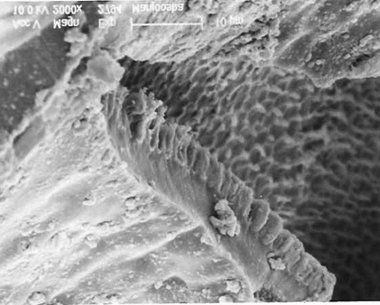
Figure 7 Cellular structure of seed showing micropyle and cotyledons. Cu, cuticle; Ep, epidermis; Oi, outer integument; Ii, inner integument; Hc, hourglass cells; Vr, vascular regions; Ed, endosperm; Cot, cotyledons; Pc, palisade-like cells; Ms, mesophyll; Ie, inner epidermis.
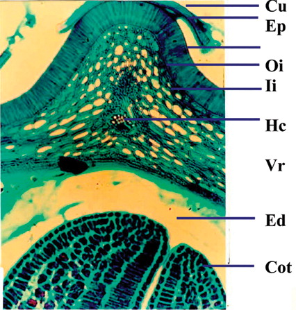
Figure 8 Cellular structure of seed showing seed coat and endosperm. Cu, cuticle; Ep, epidermis; Oi, outer integument; Ii, inner integument; Hc, hourglass cells; Vr, vascular regions; Ed, endosperm; Cot, cotyledons; Pc, palisade-like cells; Ms, mesophyll; Ie, inner epidermis.
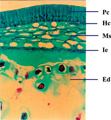
Figure 9 Cellular structure of cotyledon.
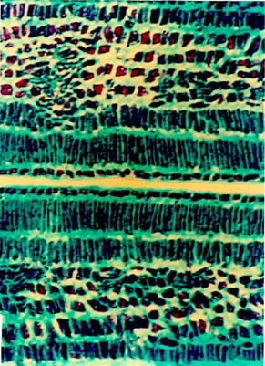
Figure 10 Endospermal region in T.S. of seed.
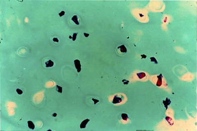
Table 1 Powder studies in seed of C. angustifolia..
Figure 11 Physicochemical parameters of Cassia angustifolia. seeds.
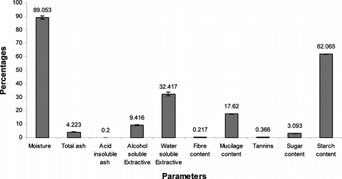
Figure 12 Successive extractive values of Cassia angustifolia. seeds.
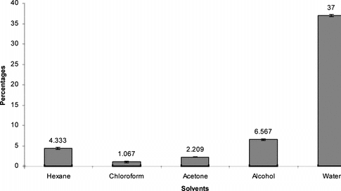
Figure 13 HPTLC fingerprint profile of different samples of Cassia angustifolia. seeds procured from different regions (1–4) along with the active constituents sennoside A and B.
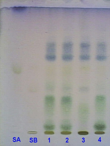
Table 2 Percentage concentration of sennoside A and B in seed samples of different geographic regions.