
Figures & data
Figure 1. Diaphragm. A high-frequency linear array probe is utilized to identify the diaphragm located between the eighth to tenth intercostal space. The resulting image typically displays three parallel high and low echo bands, with the upper and lower layers representing the pleural and peritoneal membranes, respectively, and the diaphragm located in the middle.
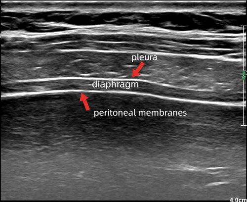
Figure 2. Diaphragm excursion. The M-mode is utilized to detect diaphragm movement. And diaphragm excursion refers to the vertical distance from the baseline to the highest point at the end of both inspiration and expiration.
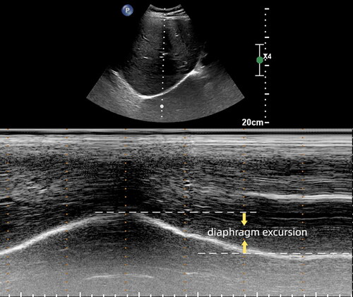
Table 1. Characteristics and findings of articles using diaphragmatic ultrasound in NIV.