Figures & data
Figure 1. DNA packaging of the related phages, T3 and T7. (A) DNA packaging in vivo is illustrated with capsid II participating in the type 1 and proposed type 2 cycles (reviewCitation4). (B) U-NLD capsid II and particles from neighboring fractions [fraction density (g/ml) indicated at the top] are analyzed by SDSPAGE in a 9% polyacrylamide separating gel. The lane labeled WT-CII has wild type T3 NLD capsid II; the lane labeled U-NLD has T3Su-1 U-NLD capsid II. The arrow indicates the direction of electrophoresis; arrowheads indicate origins of electrophoresis. Some gp9 scaffolding protein is present in capsid II. The gp9/gp10 ratio is, however, higher for capsid I. For reasons not known, wild type gp8 formed a doublet band, which it usually does not. (C) EM of U-NLD capsid II with the procedure of references Citation27 and Citation28.
![Figure 1. DNA packaging of the related phages, T3 and T7. (A) DNA packaging in vivo is illustrated with capsid II participating in the type 1 and proposed type 2 cycles (reviewCitation4). (B) U-NLD capsid II and particles from neighboring fractions [fraction density (g/ml) indicated at the top] are analyzed by SDSPAGE in a 9% polyacrylamide separating gel. The lane labeled WT-CII has wild type T3 NLD capsid II; the lane labeled U-NLD has T3Su-1 U-NLD capsid II. The arrow indicates the direction of electrophoresis; arrowheads indicate origins of electrophoresis. Some gp9 scaffolding protein is present in capsid II. The gp9/gp10 ratio is, however, higher for capsid I. For reasons not known, wild type gp8 formed a doublet band, which it usually does not. (C) EM of U-NLD capsid II with the procedure of references Citation27 and Citation28.](/cms/asset/63bc9d4e-0145-46f2-964a-6194b566eda8/kbac_a_1268664_f0001_oc.gif)
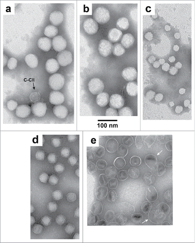
Figure 2. EM at higher magnification of (A–C) T3Su-1 U-NLD capsid II, (D) T3 heads from the study of reference Citation19, and (E) T3Su-1 NHD capsid II.