Figures & data
Table 1 The Primer Sequences
Figure 1 ED-71 increases bone mass and decreases the number of osteoclasts in vivo. (A) HE staining of rat femur in Sham, OVX, OVX+ED-71 groups at 8 weeks. Bar, 200μm. (B) The statistical analysis for the BV/TV, Tb.N, Tb.Th and Tb.Sp in the HE staining. (C) Masson staining of rat femur at 8 weeks. Bar, 200μm. (D) The immunofluorescence staining of Ctsk in rat femur at 8 weeks. Bar, 75μm. (E) The statistical analysis of the number of Ctsk-positive osteoclasts. Error bars stand for mean ± SD (n=5). *P < 0.05. **P < 0.01. ***P < 0.001. OVX, ovariectomy; OVX+ED-71, ovariectomy + eldecalcitol; GP, group plate; TB, trabeculae bone; BM, bone marrow; Ctsk, cathepsin K; BV/TV, bone volume/tissue volume; Tb.N, trabecular bone number; Tb.Th, trabecular bone thickness; Tb.Sp, trabeculae bone space; N.Oc, number of Ctsk-positive osteoclasts.
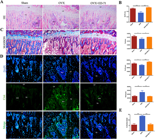
Figure 2 ED-71 activates EphrinB2-EphB4 signaling and decreases RANKL/OPG ratio. (A-D) The immunofluorescence staining of EphB4, EphrinB2, RANKL, OPG in rat femur in Sham, OVX, OVX+ED-71 groups at 8 weeks. Bar, 75μm. (E-H) The statistical analysis of fluorescence intensity of EphB4, EphrinB2, RANKL, OPG. (I) Relative ratio of RANKL to OPG in rat femur at 8 weeks. Error bars stand for mean ± SD (n=5). ***P < 0.001.
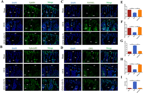
Figure 3 ED-71 promotes the expression of EphrinB2, EphB4 and OPG, and inhibits the expression of RANKL in osteoblasts. (A) Proliferation of MC3T3-E1 cells after 50 μM, 100 μM, 200 μM, 500 μM H2O2 stimulating by CCK-8 assay. (B) The mRNA expression of EphrinB2, EphB4, RANKL and OPG in MC3T3-E1 cells were detected by RT-PCR in Control, H2O2 and H2O2 + ED-71 groups after 3, 5, 7 days of induce. (C) The protein level of EphrinB2, EphB4, RANKL and OPG in MC3T3-E1 cells was detected by Western blot on the day 7 of osteogenic induction. (D) The statistical analysis of Western blot results. (E) Relative ratio of RANKL to OPG in the protein level. (F) The secretion of RANKL in MC3T3-E1 cells was detected by ELISA after 7 days of induce. (G) The immunofluorescence stanning of EphB4 in MC3T3-E1 cells on the day 7 of osteogenic induction. Bar, 50 μm. (H) The immunofluorescence stanning of EphrinB2 in MC3T3-E1 cells on the day 7 of osteogenic induction. Bar, 75 μm. (I and J) The statistical analysis of fluorescence intensity. All experiences were carried out by at least 3 times and data were expressed as mean ± SD. *P<0.05. **P<0.01. ***P<0.001. ns, no significance. H2O2 + ED-71, H2O2 + eldecalcitol.
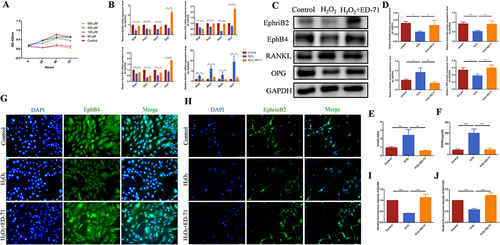
Figure 4 The inhibitory effect of ED-71 on RANKL/OPG is reversed by knocking down EphB4 in MC3T3-E1 cells. (A) The effective small interfering RNA was screened by RT-PCR. (B) The mRNA expression of EphB4, RANKL and OPG in MC3T3-E1 cells in NC, ED-71+NC, SiEphB4, and ED-71 + SiEphB4 groups was detected by RT-PCR after 7 days of induce. All groups were stimulated by H2O2. (C) The immunofluorescence staining of EphB4 in MC3T3-E1 cells after siEphB4 transfection. Bar, 100 μm. (D) The statistical analysis of fluorescence intensity. (E) The secretion of RANKL was detected by ELISA after siEphB4 transfection. (F) The protein level of EphB4, RANKL and OPG in MC3T3-E1 cells were detected by Western blot after 7 days of induce. (G) The statistical analysis of Western blot results. (H) Relative ratio of RANKL to OPG in the protein level after the siEphB4 transfection. All experiments were carried out by at least 3 times and data were expressed as mean ± SD. *P<0.05. **P<0.01. ***P<0.001. NC, negative control; ED-71+NC, eldecalcitol+ negative control; SiEphB4, small-interfering EphB4; and ED-71 + SiEphB4, eldecalcitol+ small-interfering EphB4.
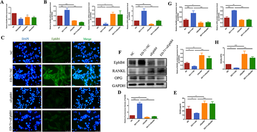
Figure 5 ED-71 inhibits osteoclasts differentiation through EphrinB2-EphB4-RANKL axis under indirect coculture conditions. (A) RAW264.7 cells were cocultured indirectly with CM containing the culture supernatant of MC3T3-E1 cells in Control, H2O2 and H2O2 + ED-71 groups for 7 days. The mRNA expression of MMP9 and Ctsk was detected by RT-PCR. (B) RAW264.7cells were cocultured indirectly with CM containing the culture supernatant of MC3T3-E1 cells in Control, H2O2 and H2O2 + ED-71 groups for 7 days. The protein level of MMP9 and Ctsk was detected by Western blot. (C) The statistical analysis of Western blot results. (D) RAW264.7 cells were cocultured indirectly with CM containing the culture supernatant for MC3T3-E1 cells in NC, ED-71+NC, SiEphB4, and ED-71 + SiEphB4 groups for 7 days. The mRNA expression of MMP9 and Ctsk in RAW264.7 cells were detected by RT-PCR. (E) The protein level of MMP9 and Ctsk in RAW264.7 cells were detected by Western blot. (F) The statistical analysis of Western blot results. All experiences were carried out by at least 3 times and data were expressed as mean ± SD.*P<0.05. **P<0.01. ***P<0.001. CM, conditioned medium; MMP9, Matrix metalloproteinase-9.
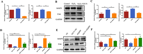
Figure 6 ED-71 activates the PI3K/AKT pathway. (A) The protein level of P-PI3K and P-AKT in MC3T3-E1 cells in Control, H2O2 and H2O2 + ED-71 groups were detected by Western blot after osteogenic induction in 7 days. (B) The statistical analysis of Western blot results of P-PI3K and P-AKT. (C and D) The immunofluorescence staining of P-PI3K and P-AKT in MC3T3-E1 cells on the 7th day of osteogenic induction. Bar, 75 μm. (E and F) The statistical analysis of fluorescence intensity. All experiences were carried out by at least 3 times and data were expressed as mean ± SD. *P<0.05. **P<0.01. ***P<0.001.
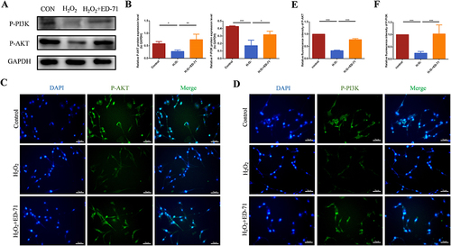
Figure 7 ED-71 decreases the RANKL/OPG ratio through the EphB4-PI3K/AKT axis. (A and B) The immunofluorescence staining of P-PI3K and P-AKT in MC3T3-E1 cells after SIEphB4 induction. Bar, 75 μm. (C and D) The statistical analysis of fluorescence intensity in MC3T3-E1 cells in NC, ED-71+NC, SiEphB4, and ED-71 + SiEphB4 groups. All groups were stimulated by H2O2 for 2 hours. (E) The protein level of P-PI3K and P-AKT in MC3T3-E1 cells were detected by Western blot. (F) The statistical analysis of Western blot results. (G) The protein level of P-PI3K, P-AKT, RANKL and OPG in MC3T3-E1 cells after being treated with LY294002(PI3K inhibitor) on the day 7 of osteogenic induction. (H) The statistical analysis of Western blot results in MC3T3-E1 cells in H2O2, H2O2+ LY294002, H2O2 + ED-71and H2O2 + ED-71+ LY294002 groups. (I) Relative ratio of RANKL to OPG in the protein level. (J) The protein level of P-AKT, RANKL and OPG in MC3T3-E1 cells after being treated with ARQ092 (AKT inhibitor) on the day 7 of osteogenic induction. (K) The statistical analysis of the P-AKT, RANKL and OPG Western blot results and relative ratio of RANKL to OPG in the protein level in MC3T3-E1 cells in H2O2, H2O2+ ARQ092, H2O2 + ED-71 and H2O2 + ED-71+ ARQ092 groups. All experiences were carried out by at least 3 times and data were expressed as mean ± SD. *P<0.05. **P<0.01. ***P<0.001. H2O2+ LY294002, H2O2+ PI3K inhibitor; H2O2 + ED-71+ LY294002, H2O2+ ED-71+PI3K inhibitor; H2O2+ ARQ092, H2O2+ AKT inhibitor; H2O2 + ED-71+ ARQ092, H2O2+ ED-71+ AKT inhibitor.
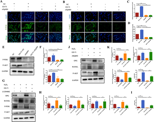
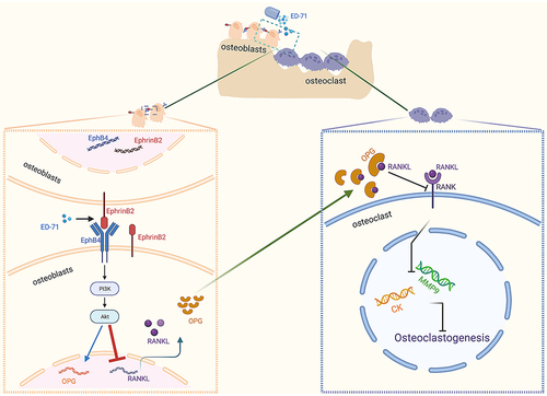
Figure 8 Pattern Diagram: ED-71 activates the PI3K/AKT pathway through EphrinB2-EphB4 signal to inhibit the expression of RANKL. Created by BioRender.com.