Figures & data
Figure 1 Anterior (A) and lateral (B) neck view of the patient with thyroid gland abscess.
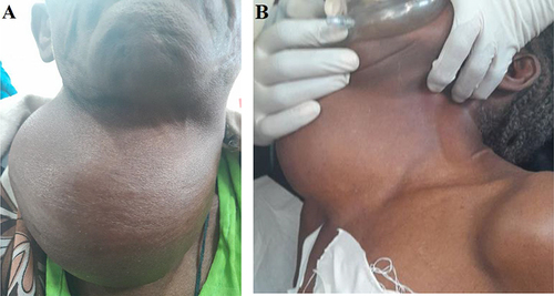
Figure 2 Rigid bronchoscopy of the patient showing nearly closed glottis before intubation.
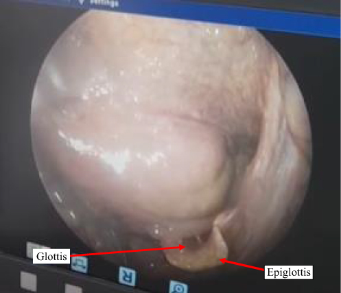
Table 1 Laboratory Results of the Patient Upon Initial Presentation and at Discharge
Figure 3 Bedside neck ultrasound of the patient showing hypoechoic lesion in the right lobe of the thyroid gland as depicted by the arrow.
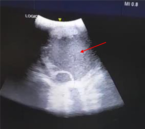
Figure 4 Head and neck post-contrast CT scan of the patient. (A) axial view of a huge thyroid abscess more on the right side as depicted by the red arrow; (B) coronal view of the same lesion causing lateral compression of the trachea and esophagus; (C) sagittal view of the same lesion.
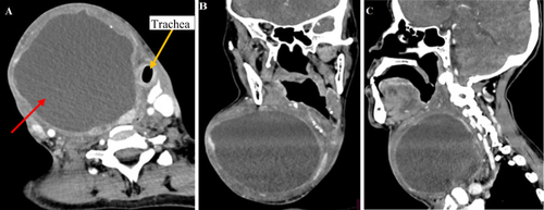
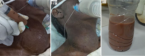
Figure 5 (A) Needle aspiration of thyroid abscess; (B) after draining 450 mL of pus; (C) drained pus.