Figures & data
Table 1 Characteristic Features of Optic Neuritis Associated with MOG-IgG, AQP4-IgG and Multiple Sclerosis (MS)
Figure 1 Continued.
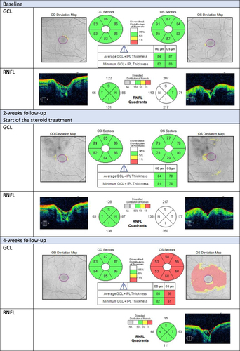
Figure 1 Evaluation of ganglion cell layer (GCL) and retinal nerve fiber layer (RNFL) in Optical Coherence Tomography (OCT) of a patient with myelin oligodendrocyte glycoprotein (MOG) antibody-associated optic neuritis during 1-year follow-up.
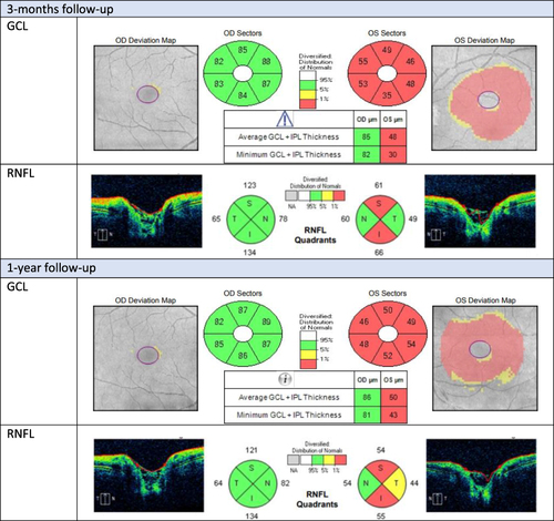
Figure 2 Evaluation of visual evoked potentials (PVEP) of a patient with myelin oligodendrocyte glycoprotein (MOG) antibody-associated optic neuritis in the left eye during 1-year follow-up.
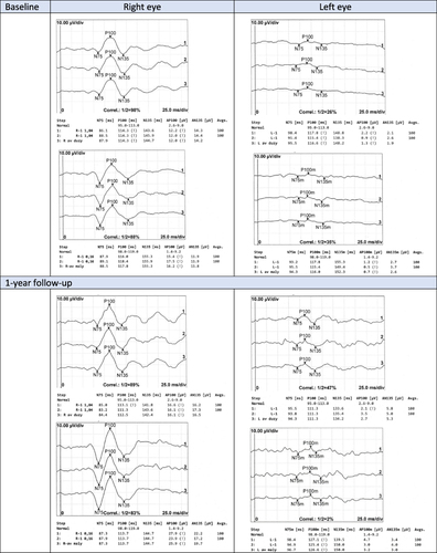
Figure 3 Evaluation of the optic nerve disc appearance of a patient with myelin oligodendrocyte glycoprotein (MOG) antibody-associated optic neuritis during 1-year follow-up.
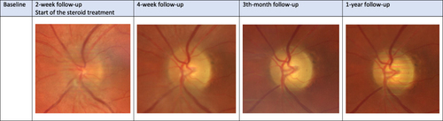
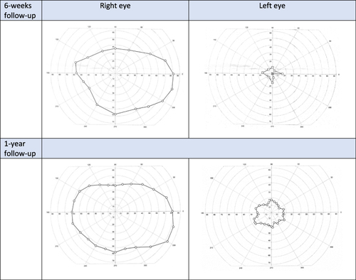
Figure 4 Evaluation of visual field of a patient with myelin oligodendrocyte glycoprotein (MOG) antibody-associated optic neuritis in the left eye during 1-year follow-up.