Figures & data
Table 1 Baseline Data and Oxygen Saturation Values of Retinal Vessels in Diabetes Mellitus Patients and Healthy Subjects
Table 2 Results of Pentacam Measurements in Diabetes Mellitus Patients and Healthy Subjects
Figure 1 Association between oxygen saturation of retinal arterioles (in %), retinal arteriovenous saturation difference (in %), and difference between central thinnest corneal thickness and peripheral thickness of the cornea measured at a concentric ring with a diameter of 4 mm (PAC Diff 4 mm, in µm) in diabetes mellitus.
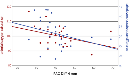
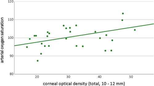
Figure 2 Correlation between retinal arterial oxygen saturation (in %) and peripheral corneal optical density (of all layers at 10 to 12 mm diameter, in µm) in diabetes mellitus patients.