Figures & data
Figure 1 CT of the abdomen and pelvis demonstrating the pelvic mass and large ascites.
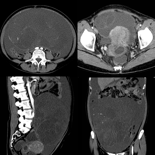
Figure 2 A ruptured 25 cm right ovarian neoplasm was found at the time of surgery.
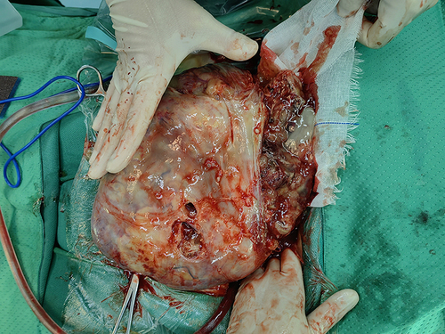
Figure 3 Immunohistochemical staining of GAS. Histologic ((A) hematoxylin and eosin staining) and immunohistochemical (B–D) findings. (A) The tumor shows gastric-type differentiation with abundant clear or pale, eosinophilic cytoplasm with atypical nuclei. (B and C) Immunohistochemical staining for MUC6 and CEA was positive. (D) P53, wild type.
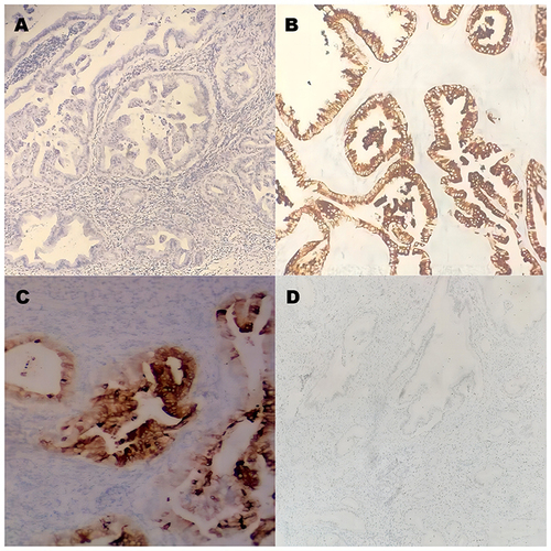
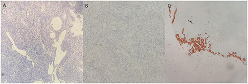
Figure 4 Immunohistochemical staining of squamous cell carcinoma (A) A high-grade squamous intraepithelial lesion and microinvasive squamous cell carcinoma were found on the cervix. (B) P53 was the wild type. (C) Immunohistochemical staining was positive for P16 in squamous cell carcinoma and negative in GAS.