
Figures & data
Figure 1 Abdominal computed tomography shows a slightly high-density heterogeneous mass of 9.3 * 5.9cm in the small intestine, with a density ranging from 34 to 57HU, indicated by yellow arrows (a). Enhanced scanning shows mild heterogeneous enhancement of the tumor, indicated by blue arrows (b).
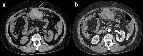
Figure 2 During the operation, a dark red mass was found on the left side of the upper abdomen, with some of the greater omentum adhering to it. It was approximately 90 cm away from the Treitz ligament and was approximately 10 * 6cm in size (yellow arrows).
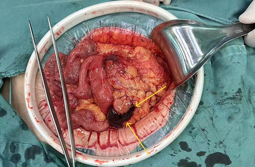
Figure 3 The lesion is located in the empty mesangium, with spindle shaped tumor cells and obvious atypia, indicated by yellow arrows (a). Tumor cells surround irregular blood vessel cavities, with green arrows (b and c). Some areas of the tumor show bleeding foci, indicated by blue arrows (d).
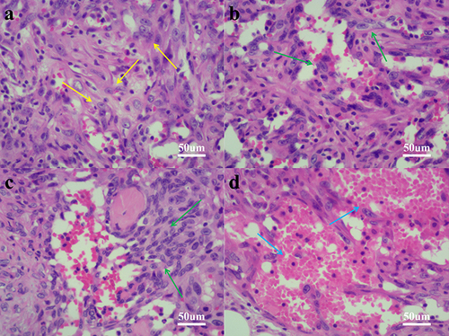
Figure 4 Immunohistochemical staining showed that tumor cells expressed CD31(a), CD34(b), Fli-1(c), MDM2(d), ERG(e), and INI1(f).
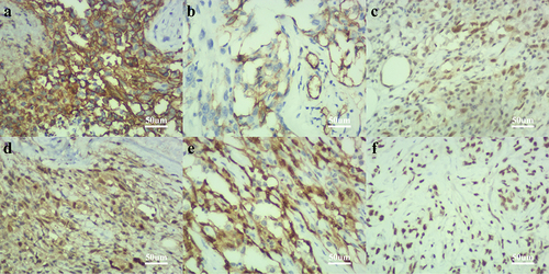
Table 1 A Retrospective Study Was Conducted on 38 Patients with Primary Gastrointestinal Angiosarcoma
Table 2 A Retrospective Study Was Conducted on 38 Patients with Primary Gastrointestinal Angiosarcoma