Figures & data
Table 1 Breast MRI: cross-over studies between gadobenate dimeglumine and other GBCAs
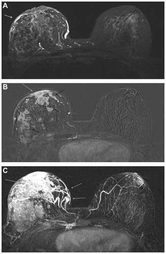
Figure 1 48-year-old woman with BI-RADS 6 lesions in the right breast detected on XR mammography and ultrasound (triple negative, IDC).
Notes: Woman underwent breast MRI with gadobenate dimeglumine (MultiHance®;) for local staging. MRI detected more nodules in the right breast than conventional imaging and allowed a better evaluation of skin thickness. It was considered multicentric disease and neoadjuvant chemotheraphy was planned. Breast MRI also revealed an enhancing lesion in the left breast, not detected on conventional imaging; second look ultrasound was performed and histological examination proved a DCIS: (A–B) IDEAL water only T2w sequence, axial plane. The grey arrow shows skin thickness while black arrows indicate a round, hyperintense nodule in the right breast. VIBRANT T1w precontrast sequence, axial plane, 3T magnet. (C) VIBRANT T1w dynamic sequence, axial plane, 3T magnet. The lesion between inferior quadrants of the right breast has a central area of necrosis (black arrow) and multiple nodules are detectable in the ipsilateral breast (grey arrow). In the left breast there is an enhancing nodule (arrowhead).
Abbreviations: MRI, magnetic resonance imagining; BI-RADS, Breast imaging-reporting and data system; IDC, invasive ductal carcinoma; XR, X-ray; DCIS, ductal carcinoma in situ.
Abbreviations: MRI, magnetic resonance imagining; BI-RADS, Breast imaging-reporting and data system; IDC, invasive ductal carcinoma; XR, X-ray; DCIS, ductal carcinoma in situ.
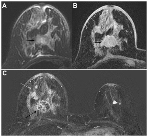
Figure 2 38-year-old woman with nipple retraction.
Notes: Ultrasound did not detect any lesions. Breast MRI with gadobenate dimeglumine (MultiHance) confirmed nipple retraction and detected non mass-like enhancement with nipple involvement. (A) T1w sequence, axial plane. Nipple retraction is due to retroareolar non-mass enhancement (white arrow). IS/T curve type II–III. The patient underwent surgery and it was proven to be IDC. (B) T2w water only sequence, nipple retraction is recognizable (white arrow).
Abbreviations: MRI, magnetic resonance imagining; IDC, invasive ductal carcinoma.
Abbreviations: MRI, magnetic resonance imagining; IDC, invasive ductal carcinoma.
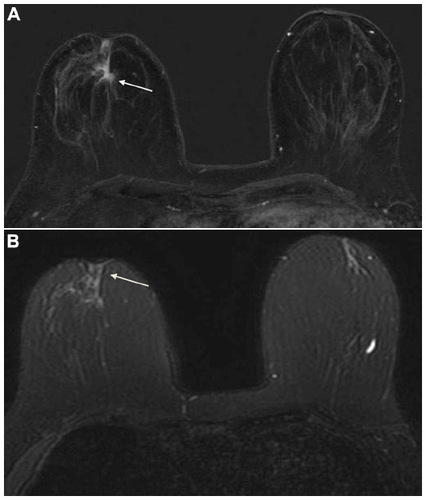
Figure 3 Breast MRI of a 59-year-old woman revealed a greater extension of the lesion on the right breast than XR mammography and ultrasound. At pathology both lesions were proved to be invasive ductal carcinoma.
Notes: T1w sequence, axial plane. Breast MRI with gadobenate dimeglumine (MultiHance) allowed detection of two lesions in both breasts. Non-mass enhancement in the right breast was previously underestimated at XR mammography and ultrasound. (A) T1w sequence after gadobenate dimeglumine (MultiHance). Non-mass enhancement (grey arrow); type III curve (B) T1w sequence after contrast agent. Mass lesion (grey arrow). Type III curve.
Abbreviations: MRI, magnetic resonance imagining; XR, X-ray.
Abbreviations: MRI, magnetic resonance imagining; XR, X-ray.
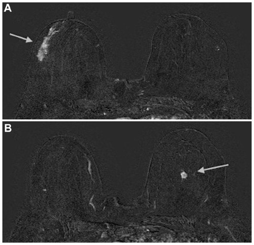
Figure 4 A 53-year-old woman with multiple lesions of the right breast, as seen on a breast ultrasound, proved to be invasive ductal carcinoma at pathology. She underwent a breast MRI to determine the extent of the disease and the multicentricity.
Notes: (A) T2w sequence, axial plane, 1.5T magnet. Asymmetry between breasts and skin thickness (grey arrow) of the right breast are recognizable. (B) T1w dynamic sequence. After gadobenate dimeglumine (Multihance) administration it is possible to detect multiple enhancing lesions (black arrows) in the right breast. Moreover skin and nipple-areola complex (grey arrow) involvement are well appreciable. IS/T curve type III. (C) Maximum intensity projection (MIP) images in the axial plane. The right breast shows increased vascularity (grey arrows).
Abbreviations: MRI, magnetic resonance imagining.
Abbreviations: MRI, magnetic resonance imagining.