Figures & data
Figure 1. FDG PET (top), PET/CT (middle) and CT (bottom) images showing a high intensity FDG uptake in a lytic-sclerotic lesion involving the body and left transverse process of T4 vertebra and the left costovertebral aspect of the adjacent 4th rib. CT, computed tomography; PET, positron emission tomography.
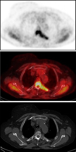
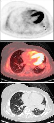
Figure 2. FDG PET (top), PET/CT (middle) and CT (bottom) images showing a very low intensity FDG uptake (arrow) in a large lung mass in the lingula. CT, computed tomography; PET, positron emission tomography.