Figures & data
Figure 1. Scheme of the filtration process for the production of PL-PPC. Whole blood-derived PL concentrates passing quarantine were thawed, centrifuged, pooled in a 5-L bag and pre-filtered through a filter with pore sizes of 0.80/0.45 μm and sterile filtered into a second 5-L bag. In a second step, the final filtration using a filter with pore sizes of 0.45/0.35 μm was performed into a third 5-L bag and filled into containers of suitable sizes.
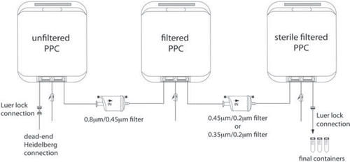
Figure 2. Multiplex analysis of human PL. (A) Cytokine concentrations of different PL-PPC preparations in pg/mL. Number of analyzed batches: n = 3 for G-CSF, GM-CSF, INF-γ, TNF-α, IL-1α, IL-1β, IL-2, IL-6, IL-7, IL-8, IL-10, VEGF, MIP-1α, MIP-1β, GRO (CXCL1/2/3), TGF-β1 and bFGF; and n = 9 for sCD40L, sVCAM-1, sICAM-1, PDGF-AA, PDGF-AB/BB and RANTES. (B) Comparing cytokine content of PL-PPC and PL-APC. Number of analyzed batches: n = 3 for TGF-β1 and bFGF in PL-APC and PL-PPC; n = 4 for sCD40L, sVCAM-1, sICAM-1, PDGF-AA, PDGF-AB/BB and RANTES in PL-APC; and n = 6 for these cytokines in PL-PPC. Open bars depict PL-PPC and filled bars PL-APC.
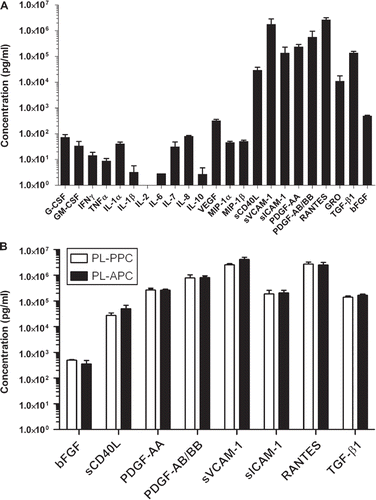
Figure 3. Stability of cytokines. (A) Human cytokine analysis of one PL-PPC batch stored at + 4°C. Starting just after production, the concentration of six highly present soluble factors was evaluated by multiplex cytokine dosage every week for 4 weeks. (B) Human cytokine analysis of one PL-PPC batch incubated at + 37°C. Starting just after production, the concentration of six highly present soluble factors was evaluated by multiplex cytokine dosage every week for 4 weeks.
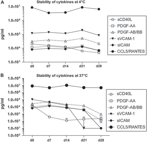
Figure 4. Proliferation capacity of PL-PPC and PL-APC. (A) Comparison of MSC proliferation in 10% PL-APC (n = 4) and 10% PL-PPC (n = 6). (B) MSC proliferation assays were performed by comparing different batches of PL-PPC (n = 6) and PL-APC (n = 2) as a culture supplement at concentrations ranging from 0 to 20%. Results are expressed as mean and SD values of three independent experiments. RFU, relative fluorescence units.
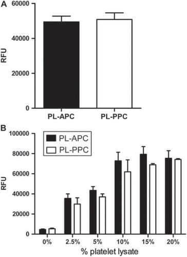
Table I. Analysis of growth-promoting effects of PL-PPC and PL-APC. Six batches of PL-PPC and three batches of PL-APC were used to compare MSC proliferation and clonogenicity parameters in T-25 culture flasks according to a GMP-grade protocol.
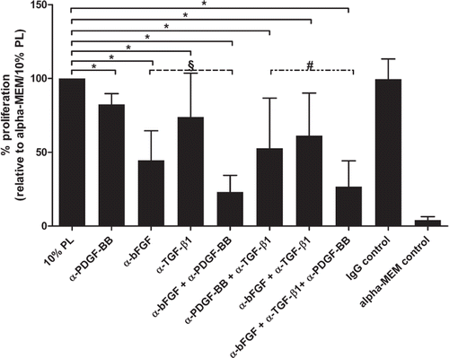
Table II. Comparison of growth-promoting effects of PL-PPC and FBS in T-25 culture flasks.
Table III. Comparison of growth-promoting effects of PL-PPC and PL-APC in a 5-chamber stack system performed according to a GMP-grade standardized protocol.
Figure 5. Inhibition of PDGF-BB by a monoclonal antibody significantly reduces proliferation of MSC in medium supplemented with 10% PL (P < 0.001). Addition of recombinant human PDGF-BB (20 ng/mL) did not enhance proliferation compared with 10% PL alone (P = 0.47). Shown are mean and SD values of quadruplicate wells of one representative experiment. Each experiment was repeated at least twice. RFU, relative fluorescence units; rh, recombinant human; α, anti; n.s., not significant.
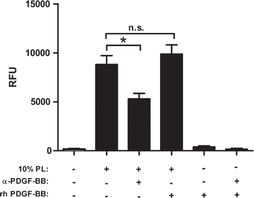
Figure 6. Inhibition of PL components bFGF, PDGF-BB and TGF-β1 by neutralizing antibodies as single factors or in combination significantly reduces MSC proliferation. Data are presented as mean and SD values of quadruplicate wells of three independent experiments. Statistical significance of the effect of inhibition of bFGF, TGF-β1 and/or PDGF-BB on MSC proliferation: *P < 0.05 compared with MSC cultivated in alpha-MEM/10% PL; #P = 0.039 compared with anti-PDGF-BB + anti-TGF-β1; §P = 0.039 compared with anti-PDGF-BB. α, anti.