
Abstract
Background: Onychopapilloma is an uncommon benign tumor of the nail bed and the distal matrix. Objectives: We aimed to investigate the clinical and pathological features of onychopapilloma in Taiwan.Materials and methods: We conducted a retrospective analysis of 12 patients with histopathologically proven onychopapilloma in a medical center in southern Taiwan from 2017 to 2023. Results: This case series consisted of 5 men and 7 women aged 29 to 38, with a mean age of 41.25 years. The clinical features were as follows: distal subungual hyperkeratosis (100%), longitudinal erythronychia (50%), longitudinal leukonychia (50 %), distal onycholysis (41%), and distal nail plate fissuring (41%). The duration of the disease varied greatly, ranging from 1 month to several years. Most patients were asymptomatic (58%), while some presented tenderness (41%). Fingernail involvement was more prevalent than toe involvement, with the thumb being the most commonly affected site. Most of the patients presented with a solitary onychopapilloma. None of the seven patients who underwent surgery and were available for follow-up experienced recurrence.Conclusions: This study highlights that longitudinal erythronychia and leukonychia emerged as the predominant clinical presentations of onychopapilloma. Furthermore, our findings suggest that surgical excision appears to be an effective method for onychopapilloma.
Introduction
Onychopapilloma is an uncommon benign tumor originating from the nail bed and distal matrix. Baran and Perrin first described the tumor as ‘localized multinucleate distal keratosis’ histopathologically in 1995 (Citation1). Subsequently, in 2000, Baran named the tumor onychopapilloma (Citation2). According to previous studies (Citation3,Citation4), the typical clinical presentation of onychopapilloma is polymorphic, including longitudinal erythronychia, longitudinal leukonychia, splinter hemorrhage and longitudinal melanonychia. Furthermore, distal subungual hyperkeratosis was observed in most cases. Onychopapilloma usually occurs in a single digit; however, a notable exception was reported in Australia, where one patient exhibited onychopapillomas that affected multiple digits (Citation5). Due to its diverse clinical presentation, onychopapilloma may occasionally resemble other worrisome medical conditions, such as melanoma or squamous cell carcinoma. Misdiagnosis of malignancy could lead to substantial anxiety and worry in the patients. Skin biopsy is reliable in differentiating onychopapilloma from other malignant diagnoses. Histology of onychopapilloma showed acanthosis, papillomatous nail bed, matrix metaplasia, and distal subungual hyperkeratosis (Citation3,Citation4), while multinucleated giant cells are uncommon. Research on onychopapilloma in Asia has been relatively limited. According to a study conducted in Korea, Kim et al. found that the onychopapilloma in Korean individuals exhibits clinical and dermoscopic characteristics that resemble those seen in Caucasian individuals (Citation6). Onychopapilloma is usually asymptomatic, but can sometimes cause tenderness or functional discomfort during daily work (Citation3, Citation6). Currently, there are no definite guidelines for the most accurate surgical method for the treatment of onychopapilloma.
To our knowledge, no prior large-scale studies on onychopapilloma have been conducted in Taiwan. This article aims to evaluate the clinical characteristics and histopathological diagnosis of onychopapilloma within the Taiwan population.
Materials and methods
We retrospectively reviewed patients diagnosed with onychopapilloma at the Department of Dermatology of Kaohsiung Chang Gung Memorial Hospital from October 1, 2017 to July 31, 2023. A total of 12 patients were included in the study, and we analyzed their sex, age, tumor location, clinical symptoms, and histopathological characteristics based on their medical records.
Of the 12 patients, 11 patients received excision surgery, while one patient only received an incisional biopsy. All patients were diagnosed with onycopapilloma with histopathology proven.
Excision surgery was performed under appropriate nerve block anesthesia. After avulsion of the nail plate, we exposed the onychopapilloma on the nail bed and then carefully removed it through a longitudinal excision procedure down to the bone. Following the removal of the onychopapilloma, the defect was sutured with non-absorbable thread, and the stitches were removed two weeks later ().
Figure 1. The longitudinal excision of the onychopapilloma and histological characteristics. (a) There is longitudinal leukonychia with onycholysis and distal fissuring on the right thumb. (b) Following the avulsion of the nail plate, the onychopapilloma was exposed on the nail bed, and we removed it via longitudinal excision. And the defect was closed with a non-absorbable thread. (c) The schematic diagram of the surgical procedure. The left side shows a presurgical nail lesion. The right side demonstrates the surgical outcome with suturing over the proximal nail fold and nail plate. (d) Papillomatosis and acanthosis of the nail bed, distal subungual hyperkeratosis with parakeratosis. (e) Matrix metaplasia with abundant eosinophilic cytoplasm in the upper part of nail bed epithelium.

The specimen and pathological reports were reviewed by experienced pathologists. In this study, we defined typical pathologic features as acanthosis, papillomatosis, distal subungual hyperkeratosis, and matrix metaplasia.
Results
Clinical findings
Among the 12 patients who participated in our study (), 5 patients (42%) were male and 7 patients (58%) were female. The mean age at the time of diagnosis was 41.25 ± 11.33 years (with a range of 24 to 61 years). The duration of the disease varied, spanning from one month to several years. Except for one patient who exhibited multiple onychopapillomas (8%) on numerous fingers and toes (see ), the remaining 11 patients presented with a solitary lesion (92%). Furthermore, most of these single lesions were located on the hands (83%). The thumb was the most frequently affected site (50%), followed by the ring finger (25%) and the middle finger (8%). Among those with single lesions, only one was located on the great toe (8%).
Figure 2. Clinical features of onychopapilloma. (a) The photos were taken of a 55-year-old man who presented with a five-year history of various nail abnormalities affecting both his fingernails and toenails. These abnormalities included multiple longitudinal leukonychia with distal subungual hyperkeratosis as well as focal onycholysis and splinter hemorrhage. (b) The fingernails showed multiple longitudinal leukonychia of the previous patient. (c) A solitary onychopapilloma presented wide longitudinal leukonychia with distal onycholysis and splinter hemorrhage on the ring finger. (d) A solitary onychopapilloma presented longitudinal erythronychia with distal onycholysis on the middle finger.
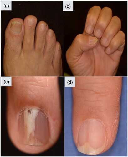
Table 1. All patients included in our study.
The clinical nail changes of onychopapilloma in this study are summarized in . The most common clinical presentations included longitudinal erythronychia (50%) and longitudinal leukonychia (50%), associated with splinter hemorrhage in 25%. (see ). Notably, no cases of longitudinal melanonychia were observed in our study. Other clinical features that were observed included distal subungual hyperkeratosis (100%), onycholysis (42%), and distal fissure (42%).
Table 2. Clinical presentation of onychopapilloma in our study compared with previous research.
Histopathologic findings
The most accurate way to diagnose onychopapilloma is by means of an oriented longitudinal excision specimen. In our study, the specimens taken from the 12 patients with onychopapilloma revealed the following histopathological features: acanthosis (100%), matrix metaplasia (100%), papillomatosis (17%), distal subungual hyperkeratosis (58%), focal parakeratosis (58%) and fibrosis of subepithelial stroma (75%) (as illustrated in and e). Furthermore, multinucleated cells were not observed in our specimens.
Our study identified typical pathological characteristics of onychopapilloma, including acanthosis, papillomatosis, distal subungual hyperkeratosis, and matrix metaplasia. Among the 12 specimens examined, 3 (25%) exhibited all of these typical findings, while the remaining 9 (75%) displayed at least two of the aforementioned pathological features.
Discussion
As far as we know, this study represents the first comprehensive analysis of the clinical and pathological features of onychopapilloma within the Taiwanese population. Our research demonstrates that longitudinal erythronychia and leukonychia are the predominant clinical manifestations of onychopapilloma. Additionally, it emphasizes the significant pathological characteristics of onychopapilloma, encompassing acanthosis, matrix metaplasia, papillomatosis, and distal subungual hyperkeratosis.
The precise cause of onychopapilloma remains unknown and three potential hypotheses have been proposed for its origin (Citation9): (i) neoplastic hyperplasia of the epithelium in the nail bed, (ii) reactive hyperplasia in response to chronic irritation or injury, and (iii) a concomitant response that occurs along with other inflammatory nail diseases. Consistently, in various studies, including our own, it has been observed that the thumb is the most frequently affected digit. This suggests that trauma or chronic irritation may play a significant role in the development of onychopapilloma, as the thumbs are more vulnerable to injury in daily life.
In our study, the prevalence of men (41.7%) was higher than that reported in studies conducted in Western countries, such as those by Tosti et al. (29.9%) (Citation6). However, the prevalence was less than that observed in another Asian study conducted in Korea by Kim et al. (59.0%) (Citation6). The average age of patients at the time of diagnosis in our study was 41.25 ± 11.33 years, which is younger than the average age reported in the study by Kim et al. (46.1 years) (Citation6) and Delvaux et al. (46 years) (Citation4). The thumb exhibited the highest incidence among all the digits, aligning with the findings of previous studies.
The most prevalent nail abnormalities observed in cases of onychopapilloma in our study were longitudinal erythronychia (50.0%) and longitudinal leukonychia (50.0%). Longitudinal erythronychia may initiate from localized damage to the nail matrix caused by onychopapilloma (Citation8). This phenomenon leads to the thinning of the nail plate and the formation of a longitudinal groove along the ventral surface of the nail plate.Moreover, the thinning of the nail plate facilitates the visibility of underlying erythema. This sequential progression ultimately contributes to the manifestation of longitudinal erythronychia in cases of onychopapilloma.
Onychopapilloma is one of the most common causes of single-localized longitudinal erythronychia. Other important differential diagnoses include glomus tumors, Bowen’s disease, warts, and melanoma (Citation8). The subungual glomus tumor is characterized by a history of pain induced by factors such as transient ischemia, exposure to cold or even light touch (Citation10,Citation11). Bowen’s disease, a form of squamous cell carcinoma in situ, may present as periungual or subungual verrucous tumors. It often manifests without significant symptoms (Citation12,Citation13). And, Bowen’s disease typically not present distal subungual hyperkeratosis, which is a common feature in onychopapilloma. Subungual warts may also present as a keratotic mass in the distal subungual area, similar to onychopapilloma. However, subungual warts can be distinguished by the presence of dotted dilated vessels when observed by dermoscopy. Moreover, the common underlying causes of polydactylous longitudinal erythronychia encompass lichen planus, Darier disease, primary amyloidosis, or graft-versus-host disease. In the cases of Darier disease and lichen planus, nail manifestations mainly occur as multiple longitudinal erythronychia (Citation14–16). Additional cutaneous findings of Darier disease, such as scaly and greasy papules in seborrheic areas and cutaneous features of lichen planus, including flat-topped polygonal papules or oral involvement, may aid in differential diagnosis. In the aforementioned medical conditions, polydactylous longitudinal erythronychia can be the only noticeable clinical manifestation at times. In such cases, it is advisable to perform a thorough and comprehensive evaluation to establish a diagnosis or identify any underlying conditions. It is worth noting that while onychopapilloma presenting with polydactylous longitudinal erythronychia has been relatively rare in previous studies, it is not impossible. For example, a case of an Australian patient with multiple digit onychopapillomas was previously reported (Citation5).
Furthermore, our research observed a higher incidence of longitudinal leukonychia (50.0%) compared to previous studies (Citation3–7). Longitudinal leukonychia in onychopapilloma is suspected to result from altered light refraction and fibrosis of the nail bed stroma, which is associated with metaplasia of the nail bed epithelium.
Longitudinal melanonychia was not observed in any of our cases of onychopapilloma. In previous studies, melanonychia can manifest as the sole presentation of onychopapilloma or in combination with other clinical characteristics (Citation17,Citation18). Onychopapilloma located in the distal nail matrix can stimulate adjacent melanocytes, leading to the development of longitudinal melanonychia. Clinically, onychopapilloma with longitudinal melanonychia can mimic nevus, Bowen disease, SCC, and melanoma. Misdiagnosis in such cases could cause patient distress and anxiety.
In this study, all patients exhibited distal subungual hyperkeratosis. This distinctive presentation manifested as a threadlike papule, and its features became more apparent when examined with the assistance of dermoscopy.
Both onycholysis and distal fissuring were observed in 5 (42%) patients. These findings can be attributed to the distal growth of the weakened and thinned nail plate, making it more susceptible to the impacts of daily activities. In addition, splinter hemorrhages were detected in 3 (25%) patients, which can be attributed to the nail bed being congested and trapped within the ventral groove, making it prone to damage from external trauma.
In our case series, seven patients (58%) were asymptomatic, while five patients (41%) reported experiencing tenderness. These findings are in line with a study conducted by Kim et al. (Citation6), where 23 patients (59.0%) were asymptomatic, nine patients reported tenderness (23.1%), and 15 patients (38.5%) complained of functional discomfort without tenderness. However, in the study by Delvaux et al. only 14.8% of the patients were asymptomatic (Citation4). Notably, in previous studies, nail discomfort or tenderness was often the primary reason for surgical removal. The lower proportion of asymptomatic patients in some studies may be attributed to the fact that asymptomatic individuals are less inclined to seek medical advice or intervention.
Among the 12 biopsy specimens, only three samples (25%) simultaneously displayed all the typical pathological findings, including acanthosis, papillomatosis, distal subungual hyperkeratosis, and matrix metaplasia. However, the remaining nine samples (75%) exhibited two or more of these typical pathological findings. Specifically, all samples showed acanthosis and matrix metaplasia, while seven samples (58%) revealed distal subungual hyperkeratosis and only two samples (17%) showed papillomatosis. Other prominent pathological features included focal parakeratosis (58%) and fibrosis of the subepithelial stroma (75%).
Identifying typical pathological findings can pose challenges when the nail bed epithelium is partially lost. Given the onychogenic nature of the onychopapilloma, complete separation of the tumor from the overlying nail plate was difficult. In several cases in our study, papillomatosis or acanthosis was observed in the epithelium of the nail bed beneath the removed nail plate.
At present, there are no established guidelines regarding the optimal surgical approach for treating onychopapilloma. Nevertheless, classical longitudinal excision could ensure the eradication of onychopapilloma in most conditions. Significantly, this approach has been associated with a lower recurrence rate compared to tangential longitudinal excision, which only removes superficial specimens (Citation4). To enhance the biopsy yield rate, we suggest sending both the nail bed and the nail plate above it for histopathology examination. However, it is important to note that, after partial nail removal, patients may experience abnormalities in the appearance of their nails, along with sensations of irritation. Furthermore, there is an increased risk of bleeding or discomfort at the surgical site without the cover of the nail plate.
We were able to contact seven patients after the removal surgery, and during an average follow-up period of 38 months, no recurrence of the lesion was detected. In contrast, in the study by Delvaux et al. (Citation4), a recurrence rate of 20% was observed over an average follow-up period of 50 months. In the study conducted by Tosti et al. (Citation3), they did not report any recurrence within a relatively short follow-up time of 6 months. However, the rate of recurrence may increase with longer follow-up durations.
There are some limitations in our study, including the retrospective design, lack of control groups, a relatively small sample size, and the fact that it was carried out within a single tertiary referral center. Additionally, some patients were lost to follow-up, which precluded us from assessing the potential recurrence of the nail lesion.
Our research revealed that onychopapilloma in Taiwan shares clinical and histopathological characteristics consistent with findings in Caucasian and Korean populations (Citation6). The most prevalent clinical presentations in our study were subungual hyperkeratosis, longitudinal erythronychia, and leukonychia. Surgical intervention yielded favorable treatment outcomes with a relatively low recurrence rate.
Disclosure statement
No potential conflict of interest was reported by the author(s).
Additional information
Funding
References
- Baran R, Perrin C. Localized multinucleate distal subungual keratosis. Br J Dermatol. 1995;133(1):1–6. 133 doi: 10.1111/j.1365-2133.1995.tb02496.x.
- Baran R, Perrin C. Longitudinal erythronychia with distal subungual keratosis: onychopapilloma of the nail bed and bowen’s disease. Br J Dermatol. 2000;143(1):132–135. doi: 10.1046/j.1365-2133.2000.03602.x.
- Tosti A, Schneider SL, Ramirez-Quizon MN, et al. Clinical, dermoscopic, and pathologic features of onychopapilloma: a review of 47 cases. J Am Acad Dermatol. 2016;74(3):521–526. doi: 10.1016/j.jaad.2015.08.053.
- Delvaux C, Richert B, Lecerf P, et al. Onychopapillomas: a 68-case series to determine best surgical procedure and histologic sectioning. J Eur Acad Dermatol Venereol. 2018;32(11):2025–2030. doi: 10.1111/jdv.15037.
- Yun JSW, et al. Clinical and histopathological features of onychopapilloma in an Australian setting: a case series of 50 patients. Australas J Dermatol. 2022;63(4):e350. e355.
- Kim T-R, Bae K-N, Son J-H, et al. Onychopapilloma: its clinical, dermoscopic and pathologic features. J Eur Acad Dermatol Venereol. 2022;36(11):2235–2240. doi: 10.1111/jdv.18461.
- Starace M, Alessandrini A, Ferrari T, et al. Clinical and onychoscopic features of histopathologically proven onychopapillomas and literature update. J Cutan Pathol. 2022;49(2):147–152. doi: 10.1111/cup.14119.
- Jellinek NJ. Longitudinal erythronychia: suggestions for evaluation and management. J Am Acad Dermatol. 2011;64(1):167.e1-11–167.11. doi: 10.1016/j.jaad.2009.10.047.
- Kim M, Sun EY, Jung HY, et al. Onychopapilloma: a report of three cases presenting with various longitudinal chromonychia. Ann Dermatol. 2016;28(5):655–657. doi: 10.5021/ad.2016.28.5.655.
- Netscher DT, Aburto J, Koepplinger M. Subungual glomus tumor. J Hand Surg Am. 2012;37(4):821–823; quiz 824. doi: 10.1016/j.jhsa.2011.10.026.
- Grover C, Jayasree P, Kaliyadan F. Clinical and onychoscopic characteristics of subungual glomus tumor: a cross-sectional study. Int J Dermatol. 2021;60(6):693–702. doi: 10.1111/ijd.15358.
- Wollina U. Bowen’s disease of the nail apparatus: a series of 8 patients and a literature review. Wien Med Wochenschr. 2015;165(19-20):401–405. doi: 10.1007/s10354-015-0383-4.
- Perruchoud DL, Varonier C, Haneke E, et al. Bowen disease of the nail unit: a retrospective study of 12 cases and their association with human papillomaviruses. J Eur Acad Dermatol Venereol. 2016;30(9):1503–1506. doi: 10.1111/jdv.13654.
- Cooper SM, Burge SM. Darier’s disease: epidemiology, pathophysiology, and management. Am J Clin Dermatol. 2003;4(2):97–105. doi: 10.2165/00128071-200304020-00003.
- Goettmann S, Zaraa I, Moulonguet I. Nail lichen planus: epidemiological, clinical, pathological, therapeutic and prognosis study of 67 cases. J Eur Acad Dermatol Venereol. 2012;26(10):1304–1309. doi: 10.1111/j.1468-3083.2011.04288.x.
- Jellinek NJ, Lipner SR. Longitudinal erythronychia: retrospective single-Center study evaluating differential diagnosis and the likelihood of malignancy. Dermatol Surg. 2016;42(3):310–319. doi: 10.1097/DSS.0000000000000594.
- Ito T, Uchi H, Yamada Y, et al. Onychopapilloma manifesting longitudinal melanonychia: a mimic of subungual malignancy. J Dermatol. 2015;42(12):1199–1201. doi: 10.1111/1346-8138.13097.
- Hashimoto H, Ito T, Yamada Y, et al. Onychopapilloma presenting as longitudinal melanonychia: a case report and literature review. Australas J Dermatol. 2021;62(2):244–246. doi: 10.1111/ajd.13543.