
Abstract
Because cigarette smoke can induce COPD/emphysema through accelerating senescence with or without an incomplete repair system. However, the pathogenesis of COPD following lung senescence induced by CS is not fully understood. Airspace enlargement and airway epithelial cell senescence are common finding during the COPD development. We investigated the lung tress response to CS and demonstrated that a stress-responsive transcription factor, FOXO3, was regulated by deacetylase. SIRT1 inhibited FOXO3 acetylation and FOXO3 degradation, leading to FOXO3 accumulation and activation in airway epithelial cells. CS exposure activated SIRT1 contributed to FOXO3 activation and functioned to protect lungs, as deletion of SIRT1 decreased CS-induced FOXO3 activation and resulted in more severe airway epithelial cells senescence airspace enlargement. Strikingly, deletion of FOXO3 during the development of COPD aggravated lung structural and functional damage, leading to a much more profound COPD phenotype. We show that deletion of FOXO3 resulted in decreased autophagic response and increased senescence, which may explain lung protection by FOXO3. Our study indicates that in the COPD, stress-responsive transcription factors can be activated for adaptions to counteract senescence insults, thus attenuating COPD development.
Keywords:
Instruction
Chronic obstructive pulmonary disease (COPD) is a senescence-related disease. Premature cellular senescence significantly affects the function of lung progenitor cells, leading to depletion of the stem cell pool and impairment of lung tissue repair function [Citation1]. Cell senescence is the basic feature of cell cycle arrest in all cells under internal and external stress. Recently, studies have indicated that exposure to cigarette smoke (CS) and acceleration of cell senescence are interconnected. Exposure to CS enhances the risk for COPD by 4.9-fold and the risk for airway obstruction with respiratory failure requiring respiratory support by 3.1-fold [Citation2].
The pathophysiology of COPD caused by exposure to CS has been extensively investigated, with many studies employing animal models of lung damage caused by exposure to CS [Citation3]. Injury caused by mild levels of CS exposure activates intrinsic healing processes, which result in the repair of cell senescence but have no impact on lung function [Citation4]. When subjected to an extremely severe CS exposure, the repair mechanism cannot entirely alleviate senescence, and animals eventually develop emphysema [Citation5]. Altering the duration of exposure to CS and other harmful gases is an easy method to alter the degree to which airspace is enlarged as a result of their presence [Citation6]. For instance, CS exposure for six months leads to severe senescence, resulting in enlarged airways, deteriorated lung function, poorer exercise endurance and decreased saturation of arterial oxygen [Citation7]. Mechanisms underlying the breakdown of protein and organelles are preserved during the repair of bronchial epithelial cells when severe senescence has occurred. Cell senescence and airspace enlargement are the primary characteristics of COPD. In a previous study, we examined the pathophysiology of COPD caused by severe CS exposure at both molecular and cellular levels by a series of tests over several weeks [Citation8]. However, the cellular defence system that supports recuperation and repair requires further investigation. Given that CS exposure is a potent stress signal, autophagy may be predominantly involved in the modulation of cell senescence [Citation8, Citation9]. Chronic oxidative stress has been observed in the lungs with severe injury but incomplete repair, which is most likely attributed to cell senescence and the widening of airspaces. The two primary triggers of autophagy are oxidative stress and degradation of organelles [Citation10]. We hypothesised that autophagy, which occurs in response to oxidative stress caused by exposure to CS, is the basic mechanism of epithelial cell senescence and repair. However, in the presence of chronic oxidative stress, compensatory autophagy may not be sufficient for the recovery of the structure and function of the lungs to their original, healthy state.
This study aimed to examine the relationship between exposure to CS and autophagy. The findings indicate that CS exposure suppresses the stress-responsive transcriptional factor FOXO3, which is a key modulator of epithelial autophagy in the lungs. After the exposure of CS to the lungs, acetylation and degradation of FOXO3 are accelerated, thereby elevating the levels of FOXO3 protein, which in turn stimulates stress responses and leads to epithelial autophagy [Citation11]. In addition, deletion of FOXO3 during the progression of COPD inhibits autophagy and aggravates oxidative damage, eventually resulting in significant cellular senescence and enlargement of the airways. Overall, this study demonstrates the response of mitochondria to CS-induced mitophagy modulated by the pleiotropic transcription factor FOXO3, which ameliorates the severity of COPD.
Methods
Animal models
Mice were exposed to CS as described previously [Citation12]. Briefly, mice aged 4- 8 weeks mice were produced from C57BL/6J mice procured from Jackson Laboratories, USA. Cre-lox technology was used to produce sirtuin 1-/- (SIRT1-/-) mice by crossing B6,129-Sirt1tm1Ygu/J mice bearing loxP (locus of X over P1) sites guiding the excision of exon 4 of SIRT1 with MCK-cre-expressing B6:FVB (129S4)-Tg (Ckmm-cre) 5Khn/J and PAX7-cre-expressing Pax7tm1(cre)Mrc/J mice. Mice with the B6 strain had an abundant expression of the SIRT1 gene. Mice with the Cg-Tg (SIRT1) ASrn/J strain possessed a wild-type variant of the SIRT1 gene, which is boosted by an endogenous bacterial artificial chromosome (BAC) transgene. After 2 weeks of CS exposure, mice with floxed FOXO3 alleles (FOXO3fl/fl) were administered doxycycline to facilitate FOXO3 deletion, whereas mice who had only received the vehicle treatment were included in the control group.
Lung morphometry
At a pressure of 25-cm H2O, 1% low-melt agarose was injected into the lungs of mice that had not been lavaged. The lung samples were fixed with 4% neutral buffered paraformaldehyde (PFA) [Citation13, Citation14], desiccated, embedded in paraffin and sectioned into 4-µm-thick slices using a rotary microtome Fixed lung was dehydrated, embedded in paraffin, and sectioned into 4-μm sections using a rotary microtome (MICROM International GmbH). The midsagittal slices of the lungs were stained with haematoxylin and eosin (H&E), and the Lm of airspace of the slices was calculated using the MetaMorph software (Molecular Devices) [Citation14]. In a blinded experiment, 10 100x fields were selected randomly, and the resulting images were manually analysed. The structure of the vascular system and airways was excluded from further analysis.
Measurement of senescence-associated beta-galactosidase activity
The activity of senescence-associated beta-galactosidase (SA-β-gal) was quantified by determining the conversion rate of 4-methylumbelliferyl-β-d-galactopyranoside (MUG) to the fluorescent hydrolysis product 4-methylumbelliferone (4-MU) at a pH of 6.0 as reported in a previous study [Citation15]. Briefly, lung tissue samples were homogenised in a lysis buffer (0.25-mM PMSF, 0.5-mM benzamidine, 40-mM sodium phosphate, 40-mM citric acid and 5-mM CHAPS [pH, 6.0]) and incubated on ice for 1 h. The samples were centrifuged for 5 min at 12,000 g, and the resulting supernatant was mixed with 2× reaction buffer (10-mM β-mercaptoethanol, 300-mM NaCl, 40-mM sodium phosphate, 40-mM citric acid and 4-mM MgCl2 [pH, 6.0] with 1.7-mM MUG) and incubated in a water bath at 37 °C for 3 h. Subsequently, 50 μL of the reaction mixture was added to 500 μL of stop solution containing 400-mM sodium carbonate (pH, 11.0), and 150 μL of the sample was added to each well of a 96-well plate to measure absorbance using a SpectrumMax M5 plate reader (Molecular Devices) with the following parameters: emission wavelength, 465 nm; excitation wavelength, 360 nm; integration, 40 μs; constant gain, 46. The conversion rate of MUG to 4-MU was used to calculate the normalised SA-β-gal activity, which was then expressed as the observed fluorescence intensity divided by the milligrams of total protein in the test.
Furthermore, 6-μm thick sections of frozen lung tissues embedded in OCT were stained using a commercial kit(Cell Signalling Technology) to evaluate SA-β-gal activity. Briefly, the lung tissues were fixed with 2% formaldehyde containing 0.2% glutaraldehyde for 15 min and washed twice with PBS. Thereafter, the samples were incubated with the staining solution for 24 h at 37 °C (2-mM MgCl2, 0.15-M NaCl, 40-mM citric acid/sodium phosphate, 5-nM potassium ferricyanide, 5-nM potassium ferrocyanide and 1-mg/mL X-gal in dimethyl formamide [pH, 5.9]). Cells that were stained blue were positive for SA-β-gal.
Measurement of autophagic potential
Images (×630 magnification) depicting proximal tubules stained with lotus tetragonolobus agglutinin (LTA) were randomly chosen. A minimum of 100 cells was selected in each lung to determine the number of autophagic cells. Cells were considered autophagic if they had ≥3 red fluorescence protein (RFP) spots. Autophagic cells were divided into groups based on the number of RFP spots present in each cell: 3–5, 6–10, 11–15, 16–20, 21–30, 41–50 or >50 spots. The experiment was repeated twice for quantitative analysis.
Culture and analysis of alveolar epithelial cells
The lungs of 6–8-week-old mice were harvested, and alveolar cells were subsequently isolated for primary culture according to standard procedures [Citation16, Citation17]. The cells were cultured on chamber slides for 48 h until a confluence of 70% was attained and transfected with either Ad-GFP (Vector Biolabs, 1060) or Ad-GFP-FoxO3a (Vector Biolabs,1026) at a multiplicity of infection (MOI). Thereafter, the cells were incubated with 1% cigarette smoke extract (CSE) for 2–36 h. Transfected cells cultured in a 21% oxygen environment serves as the control group. The cells were fixed with 4% cold PFA for 10 min, and the nuclear expression of FoxO3 was quantified (Affymetrix, 19943 1LT). Thereafter, they were washed with PBS, stained with DAPI and imaged at a magnification of ×400. For cell starvation, the cells were incubated with Earle’s balanced salt solution (EBSS) for 2 h and deprived of amino acids and glucose in the presence or absence of 5-mM DMKG (MilliporeSigma, 34631-5G). Cells cultured under conventional circumstances served as the control group.
Immunostaining and image analysis
Immunostaining was performed using standard techniques [Citation18]. The detailed staining procedure and antibodies used are mentioned in the Supplementary Methods section. A Zeiss AxioObserver Z1 fluorescent microscope was used to visualise the stained sections, a digital camera was used to capture photographs and the AxioVision software was used to analyse the images.
Immunoblotting and immunoprecipitation analyses
Both tissue preparation and immunoblotting (IB) were performed as described previously [Citation19]. The Supplementary Methods section includes the comprehensive procedure and information regarding antibodies. FoxO3 was immunoprecipitated using the Catch and Release V2.0 Reversible Immunoprecipitation System (MilliporeSigma, 17-500 A), and IB was performed using a pan-hydroxyl proline antibody. Briefly, total protein was extracted using RIPA buffer, and 500 μg of cellular protein was combined with 0.1 μg of FoxO3 antibody (Cell Signalling Technology, 2497), 10 μL of affinity ligand and washing buffer. The volume of the solution was set to 500 μL per column, and the solution was incubated at ambient temperature for 1 h. Subsequently, the solution was diluted by adding 70 μL of elution buffer denaturised with 5% 2-mercaptoethanol (MilliporeSigma, M-6250). To detect hydroxylation of FoxO3, the solution was heated to 95 °C for 10 min and loaded at a concentration of 10 µL per lane.
Detection of superoxide with MitoSOX red and dihydroethidium
Alveolar epithelial cells were grown on chamber slides and incubated with 1% CSE for 1 h. Subsequently, the cells were stained with either 0.5-μM dihydroethidium (DHE) (Thermo Fisher Scientific, D11347) or 0.5-μM MitoSOX Red (MilliporeSigma, M36008) in 1% CSE for 30 min, washed with PBS and sealed with coverslips. To determine the degree of oxidative stress in the lungs, 10-µm-thick freshly snap-frozen sections were incubated with 1-μM DHE in a humidity-controlled chamber at 37 °C for 10 min, washed with PBS and mounted using Vectashield (Vector, H-1200-10). The sections were promptly visualised at ×200 magnification, imaged and analysed.
Statistical analysis
The number of cells or animals used in each experiment is specified. All cell experiments were performed in duplicate, and data were expressed as mean ± SEM. Significant differences between two groups were assessed using the two-tailed Student’s t-test. A nonparametric t-test (Wilcoxon/Mann–Whitney U test) was used to compare tubular dilatation, renal injury scores and renal fibrosis (Masson’s trichrome staining) in WT and mutant mice. Differences across more than two groups were evaluated using one-way analysis of variance (ANOVA), followed by Dunnett’s post hoc test. A P-value of <0.05 was considered significant.
Result
Cigarette smoke extract transiently induced the transcriptional activity of FOXO3
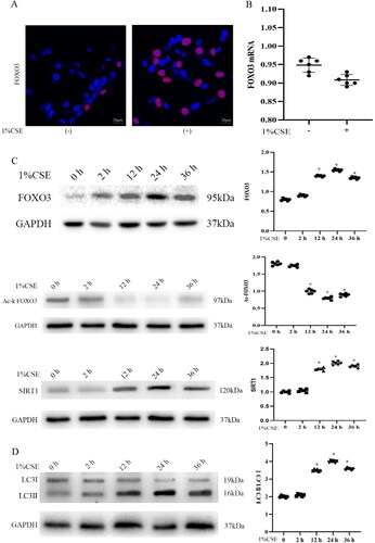
To examine the role of CSE in triggering autophagy via activation of the stress-responsive transcriptional factor FOXO3, FOXO3 was localised in the nucleus, where it performs its function. To elucidate the function of stress in activating FOXO3, primary cultures of bronchial epithelial cells were prepared in a medium designed to increase cell growth. Immunofluorescence staining for endogenous FOXO3 revealed abundant nuclear FOXO3 in cells treated with 1% CSE (); however, the levels of FOXO3 mRNA were not different between the NG and CSE groups (). The level of endogenous FOXO3 protein was considerably increased in response to stress for a period of 2–36 h (). These results indicate that stress may be responsible for the post-translational modulation of FOXO3. Deacetylation of acetylated FOXO3 by histone deacetylases (HDACs) is one of the most extensively investigated post-translational modifications of FOXO3. It is critical for reducing the translocation of acetylated FOXO3 from the nucleus to the cytoplasm, which is the site for its degradation [Citation20]. In this study, an increase was observed in the amount of nuclear FOXO3 protein, followed by a reduction in the amount of Ac-K (). Recent studies have reported that SIRT1–7 are members of the HDAC class III family, which is a group of enzymes that rely on coenzyme NDA + for hydrolysis and release nicotinamide and deacellysine. In this study, transient CSE exposure was found to significantly affect SIRT1 levels, indicating that the accumulation of nuclear FOXO3 after CSE exposure is most likely a result of decreased FOXO3 nuclear exportation and the reduction in Ac-FOXO3 levels can be attributed to an alteration in SIRT1 levels (). According to the findings of our previous study and other studies, SIRT1-mediated FOXO pathways promote autophagy, thereby protecting against apoptosis. Given that oxidative stress is a powerful mediator of autophagy, the ratio of LC3II/LC3I was examined. The ratio was increased in response to stress for a period of 2–36 h (), indicating that CSE-induced mitophagy was transient, which is consistent with the results of previous studies.
Figure 1. Cigarette smoke extract (CSE) transiently induced the transcriptional activity of FOXO3 and activated PINK1–Parkin-mediated mitophagy in HBECs. (A) Immunostaining of endogenous FOXO3 (performed without upregulation) revealed abundant nuclear FOXO3 (red) expression in cells treated with 1% CSE for 36 h. (B) Exposure of primary culture of bronchial epithelial cells isolated from 3-week-old wild-type mice to 1% CSE for 36 h did not result in changes in the levels of FOXO3 mRNA as determined via real-time RT-PCR. Cells were isolated from 6 mice, and experiments were performed in duplicate. (C) Expression level of endogenous FOXO3 and SIRT1 protein was elevated in primary cultures after exposure to 1% CSE; however, Ac-FOXO3 protein levels were decreased (n = 6 with duplicates). Cells without exposure to 1% CSE served as the control group (*p < 0.05 versus the control group). (D) Increased lung autophagy with a higher LC3II/LC3I ratio in cells treated with 1% CSE. GAPHD was used as the loading control in C and D. DAPI (blue) was used to counterstain the nuclei in A (scale bar = 20 μm). Two-tailed Student’s t-test was performed for A. One-way ANOVA followed by Dunnett’s post hoc test for multiple comparisons was conducted for C and D.
SIRT1 contributes to FOXO3 activation and COPD protection
The involvement of SIRT1 in the activation of FOXO3 in emphysema was examined. The expression levels of SIRT1 protein were elevated in 2 weeks, but decreased after 6 months of CS exposure (). These findings indicate that CS induces a transitory increase in the ratio of SIRT1-to-FOXO3 protein in alveolar epithelial cells, and these proteins play an essential role in the adaptive process that occurs in response to oxidative stress through transcriptional activation of genes that decrease cell senescence. To determine whether SIRT1 is required for CS to cause FOXO3 activation, mice with either heterozygous deletion of SIRT1 or SIRT1 overexpression were exposed to CS. Cells with SIRT1 deletion and control cells were named SIRT1-/- and SIRT1ctl, respectively. The lungs of mice that had not been exposed to CS were used to prepare primary culture cells. The levels of SIRT1 were negligible in both SIRT1-/- and SIRT1ctl untreated cells. However, after the cells were treated with 1% CSE for 6 h, SIRT1 protein was significantly accumulated in SIRT1ctl cells and was present in detectable quantity in SIRT1-/- cells. Furthermore, FOXO3 protein was present in untreated cells, and its levels were further lowered in cells either with or without SIRT1 deletion treated with 1% CSE; however, relatively low levels of FOXO3 were observed in cells with SIRT1 deletion (). These results indicate that transient CSE modulates the levels of FOXO3 protein without the involvement of SIRT1 protein; however, SIRT1 is implicated in the regulation of this process. Because there is no evidence to suggest the interaction between SIRT1 and FOXO3 at the protein level, FOXO3 mRNA was further examined. The levels of FOXO3 mRNA were elevated in control cells treated with 1% CSE for 5 and 15 min but did not increase when the cells were treated for an additional 30 min. The effects of SIRT1 deletion on the CSE-induced increase in FOXO3 mRNA were diminished (). These results indicate a stimulatory effect of SIRT1 on FOXO3 at the transcriptional level.
Figure 2. SIRT1 activates FOXO3 and protects against COPD. (A) SIRT1 protein levels were increased during COPD development (n = 4). (B) Elevation in the level of FOXO3 protein caused by exposure to CS was partly dependent on SIRT1 in primary cultures (n = 5 with duplicates) (^^p < 0.01 and *p < 0.05, SIRT1-/- versus SIRT1ctl at 0 and 6 h, respectively). Vehicle-treated mice (SIRT1ctl) served as the control group. (C) Loss of SIRT1 led to an ineffective response to 1% CSE treatment as indicated by an elevation in the mRNA expression level of FOXO3 (n = 3–8 with duplicates) (**p < 0.01 and ***p < 0.001 compared to cells not subjected to 1% CSE treatment n the SIRT1ctl or SIRT1-/- group). (D) Elevation in the nuclear expression of FOXO3 (green) in the alveolar epithelium (LTA in red) of the lungs of SIRT1 Tg mice 6 months after CS exposure (n = 4) (**p < 0.01). (E) SIRT1 loss increased SA-β-gal activity in mice exposed to CS for 6 months; however, the activity was not detected in the lungs of SIRT1 Tg mice. (F) Blots demonstrating reduced FOXO3 protein levels and LC3II/LC3I ratio in the lungs of SIRT1-/- mice compared to those of SIRT1ctl mice (n = 6) (*p < 0.05 and **p < 0.01, SIRT1-/- versus SIRT1ctl) (scale bar = 20 μm for D and 100 μm E). Statistical significance was determined via two-tailed Student’s t-test (B, D, E, and F) and one-way ANOVA followed by Dunnett’s post hoc test for multiple comparisons (C).
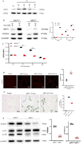
On assessing the early stages of repair, it was observed that SIRT1 protected the lungs against oxidative damage. Furthermore, to examine the role of SIRT1 in the development of COPD, mice were subjected to CS for 6 months to prevent mortality and increase the likelihood of quantifying senescence in surviving lung tissues. After 2 weeks of exposure, loss of SIRT1 led to an undetectable expression of nuclear FOXO3 in the lungs. These findings are consistent with those of the cell culture experiments. On the contrary, an increase in SIRT1 expression was associated with a reduction in nuclear FOXO3 levels, providing additional evidence that SIRT1 is implicated in FOXO3 modulation (). Compared to control mice, SIRT1 Tg mice were found to have attenuated SA-β-gal activity in their lungs after exposure to CS for 6 months (). After 2 weeks of CS exposure, IB revealed that the levels of FOXO3 protein in the lungs of SIRT1-/- mice were substantially lower than in those of control mice. In addition, the ratio of LC3II-to-LC3I was lower as a result of SIRT1 deletion (). These results suggest that SIRT1 is involved in the latter stages of lung repair following CS exposure and autophagy may be responsible for the protective effects of SIRT1 on the lungs.
FOXO3 deletion accelerates COPD development
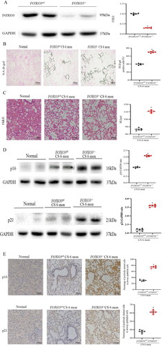
Mice with floxed alleles of FOXO3 (FOXO3fl/fl) were crossed with normal mice to examine mechanisms underlying the activation of FOXO3 in the progression of COPD. After 2 weeks of CS exposure, mice were administered doxycycline to eliminate FOXO3. Mice with and without FOXO3 deletion were named FOXO3-/- and FOXO3ctlmice, respectively. Insufficient blood supply to the lungs after ischaemia might have resulted in the partial deletion, which interfered with the delivery of doxycycline required to trigger Cre recombination. After 6 months of CS exposure, FOXO3 deletion considerably enhanced the senescent state of the lungs, indicating a serious worsening of the condition, which was verified via SA-β-gal staining (). The results of staining revealed that the airspace was enlarged, many alveoli were merged and the mean linear intercept (MLI) was increased (). Immunohistochemical (IHC) staining and western blotting of p16 and p21 (senescence-associated cyclin-dependent kinase inhibitors) were performed to verify that airway epithelial cells had reached senescence. Epithelial cells in the airways and alveoli of FOXO3-/- mice exposed to CS were positively stained for p21 and p16. In addition, western blotting revealed that the protein expression level of p21 and p16 was significantly higher in the lungs of CS-exposed FOXO3-/- mice than in CS-exposed FOXO3ctl mice (). Altogether, these results suggest that FOXO3 aggravates the severity of alveolar epithelial cell senescence, which in turn decreases the number of alveoli.
Figure 3. FOXO3 deletion accelerates COPD development. (A) Decrease in FOXO3 protein concentration in lung lysates (n = 6). (B) More severe airway epithelial cell senescence was discovered in FOXO3-/- mice than in FOXO3ctl mice. (SA-β-gal staining, n = 5) (scale bar= 100 μm). (C) Loss of FOXO3 contributed to the enlargement of the airspace caused by CS exposure for 6 months (scale bar = 100 μm). (D) Using anti-p16 and anti-p21 antibodies, western blotting was performed using lung homogenates from healthy mice and mice exposed to CS for 6 months (FOXO3-/-, FOXO3ctl). The data of densitometric analysis of the results of western blotting are averaged (±SEM) and shown in the bottom panel (n = 6). (E) Immunohistochemical staining of p16 and p21 in control mice and mice exposed to CS for 6 months (FOXO3-/-, FOXO3ctl) (scale bar = 100 μm). The proportion (average ± SEM) of airway epithelial cells that were positively stained is shown in the bottom panel (*p < 0.05, FOXO3-/- versus FOXO3ctl). Statistical significance was assessed via a two-tailed Student’s t-test (A–E).
FOXO3 deletion reduces autophagic adaptation
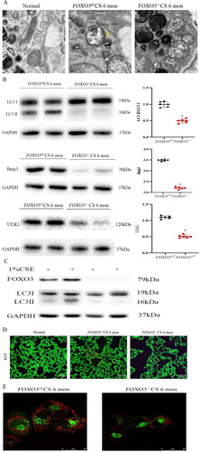
To examine whether the protective effects of unaltered FOXO3 on the lungs of mice were associated with autophagy, electron microscopy (EM) was performed, which is the industrial benchmark for identifying autophagic vesicles. After 6 months of CS exposure, autophagosomes were found to be more prevalent in the airway epithelial cells of the lungs of FOXO3ctl mice. However, FOXO3-/- mice did not have any identifiable autophagic vesicles (). Furthermore, the LC3II/LC3I ratio in the lungs of FOXO3-/- mice exposed to CS for 2 weeks was lower, indicating a decrease in autophagy. In addition, the levels of Bnip3 protein, which is both an established target of FOXO3 and an essential part of the mechanism that regulates autophagy, were reduced, and the levels of ULK protein, which plays a critical role in the beginning stages of autophagic activation, were also decreased (). To verify these in vivo results, primary cultures were prepared using cells isolated from the lung tissues that had not been treated with 1% CSE. The ratio of LC3II-to-LC3I was increased in FOXO3ctl cells, whereas that in FOXO3-/- cells did not increase significantly (). In addition, fewer RFP dots were found in airway epithelial cells with detectable FOXO3 in mice that were bred to produce FOXO3ctl and FOXO3-/- mice and subjected to CS. Autophagic spots in these airway epithelial cells were significantly reduced, resulting in an atrophic appearance of cells. However, the number of RFP dots was higher in the tubules of FOXO3ctl mice, and their airway epithelial cells were less atrophic (), indicating that FOXO3 may induce autophagy to ensure homeostatic balance in the lungs. In vivo RFP analysis revealed that primary culture cells derived from FOXO3-/- mice had fewer RFP spots than those derived from FOXO3ctl mice (). These results indicate that exposure to CS increases the autophagic capability of airway epithelial cells, which in turn enhances the activation of FOXO3.
Figure 4. FOXO3 deletion reduces autophagy. (A) EM images depicting autophagosomes (yellow arrows) in airway epithelial cells of FOXO3ctl mice 6 months after CS exposure. (B) Decreased levels of ULK1, Bnip3, LC3II and LC3I proteins were found in the lungs of FOXO3-/- mice (n = 4) (*p < 0.05, FOXO3-/- with FOXO3ctl, two-tailed Student’s t-test). (C) Primary cultures were established using cells isolated from the healthy lungs of 3-week-old FOXO3ctlmice and the lungs of FOXO3-/- mice exposed to 1% CSE for 36 h.FOXO3ctlcells demonstrated a considerable increase in LC3II/LC3I ratio compared to cells exposed to control air (p < 0.05). FOXO3-/- cells had insignificant changes in the LC3II/LC3I ratio (p = 0.31) (n = 6). Two-tailed Student’s t-test was performed for statistical analysis. (D) The autophagy reporter CREL mice were bred with FOXO3ctl and FOXO3-/- mice and exposed to CS as mentioned earlier. FOXO3-/- mice had fewer RFP dots. A considerably higher number of RFP dots was observed in the airway epithelial cells of FOXO3-/- mice (scale bar = 100 μm). (E) RFP dots in primary cultural cells indicated that FOXO3-/- cells contained fewer dots. In comparison, more RFP dots were observed in FOXO3ctl cells (scale bar = 25 μm).
FOXO3 deletion results in more mitochondrial injury
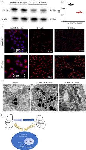
The FOXO3 gene is implicated in stress responses, and several molecules essential for the modulation of oxidative stress are among its targets. It has been demonstrated that CS enhances the production of reactive oxygen species (ROS) in lung tissues, which leads to oxidative damage. In this study, mitochondrial superoxide dismutase (SOD2), a scavenging enzyme found in mitochondria that may be induced by FOXO3 and performs a critical function in the clearance of ROS, was examined. After 6 months of CS exposure, the levels of SOD2 protein in the lungs of FOXO3-/- mice were considerably lower (). Mitochondrial superoxide levels in live cells from primary cultures isolated from FOXO3-/- mice showed a higher intensity of the fluorogenic dye MitoSOX Red compared with that seen in the FOXO3ctl cells after exposure to 1%CSE for 48 h. Additionally, the cells were incubated with DHE, a compound that undergoes oxidation when exposed to intracellular ROS and results in the generation of red fluorescence signals in the nucleus. Almost all cells isolated from the lungs of FOXO3-/- mice exhibited red nuclear signals, whereas only 36% of the cells isolated from the lungs of FOXO3ctl mice exhibited red nuclear signals. In addition, DHE staining revealed that elevated nuclear oxidative stress was observed in the lungs of FOXO3-/- mice after 6 months of CS exposure (). Furthermore, EM revealed a severe loss of cristae and enlargement of the mitochondria. In FOXO3-/- cells, many breaches were observed in both inner and outer membranes of the mitochondria (), which may result in the release of ROS into the cytoplasm, thus contributing to oxidative damage. Decreased SOD2 protein levels due to long-term chronic smoke stimulation can lead to increased mitochondrial oxidative damage, reduced autophagy, and increased cellular senescence in the lung cells of FOXO3-/- mice. Altogether, these results indicate that FOXO3 may have a preventative antioxidant impact on Pulmonary parenchyma damage. Determining the functional importance of the FOXO3/ROS pathway in the progression of COPD requires in-depth research specifically tailored to efficiently diminishing or eliminating ROS in airway epithelial cells.
Figure 5. FOXO3 deletion increases CS exposure. (A) The lungs were analysed 6 months after CE exposure. Compared to the lungs of FOXO3ctl mice, the lungs of FOXO3-/-mice had substantially lower levels of the SOD2 protein (n = 6) (*p < 0.05, FOXO3-/-versus FOXO3ctl, two-tailed Student’s t-test). (B) Left and centre panels: Primary cell cultures (FOXO3-/-or FOXO3ctl) were treated with 1% CSE for 1 h and incubated with MitoSOX Red or DHE. The levels of mitochondrial SOD2 measured via MitoSOX Red staining are shown in the left panel, indicating a higher intensity in FOXO3-/- cells. The results of DHE staining (shown in the centre panel) demonstrated that almost all (100%) FOXO3-/- cells produced red nuclear signals, whereas only 49.6% of FOXO3ctl cells generated red nuclear signals. Right panel: After 6 months of CS exposure, DHE staining revealed more pronounced nuclear oxidative stress in the lungs of FOXO3-/- mice. (C) After 6 months of CS exposure, airway epithelial cells exhibited substantial mitochondrial enlargement and loss of cristae as shown in EM images. FOXO3-/-cells had numerous ruptures in both inner and outer membranes of the mitochondria (yellow arrow) (scale bar = 10 μm for the left panel in B; 50 μm for the centre panel in B; 10,050 μm for the right panel in B; 0.5 μm for the left and centre panel in C and 0.2 μm for the right panel in C). D) Mechanistic model for FOXO3 accumulation and autophagy activation to protect lungs.
Discussion
FOXO3 is a critical downstream target of oxidative stress. The response of FOXO3a protein to stimuli is associated with autophagy, apoptosis and stress resistance. Although the involvement of FOXO members in mediating autophagy and resistance to apoptosis has been intensively studied in recent years, the role of FOXO3 activation in COPD pathogenesis, especially in protection against CSE-induced senescence via enhancement of mitophagy, remains unclear.
In this study, FOXO3 was observed to modulate adaptive responses, which counteract the harmful impact of long-term CS exposure in the lungs by increasing autophagy and reducing senescence. In contrast to the substantial cell loss that may occur following exposure to hazardous substances, chronic exposure to CS for 6 months does not contribute to a substantial increase in the shrinking and fibrosis of alveoli as a healing mechanism [Citation21]. However, autophagy, which is a process for cell survival that has been preserved throughout evolution, can be triggered by acute pulmonary CS exposure [Citation22]. Signals for the induction of autophagy can be transmitted to the sensors that monitor nutrition and energy levels, including deacetylases belonging to the sirtuin family dependent on NADH and AMPK and other molecular pathways that are implicated in stress responses [Citation16]. FOXO3 is one of the four FOXO genes found in mammals. To date, most studies have focussed on FOXO3, which regulates apoptosis, autophagy, senescence, stress responses and cell differentiation. Post-translational processes are responsible for regulating FOXO3 activity and encompass modifications such as ubiquitination, acetylation and phosphorylation [Citation23]. FOXO3 is translocated to the cytoplasm in the presence of survival factors including growth factors and nutrients. After FOXO3 is translocated to the cytoplasm, it is acetylated before it is broken down by the ubiquitin–proteasome system [Citation24, p.3]. FOXO3 is deacetylated by SIRT1 and maintained in the cytoplasm. However, FOXO3 is known to concentrate in the nucleus in response to stress, where it may subsequently operate as either a transcriptional activator or a transcriptional repressor to modulate adaptive responses [Citation5, Citation25].
The SIRT1 protein is essential for both short- and long-term adaptations that occur in response to oxidative stress. In this study, FOXO3 stimulation was confirmed to be partly dependent on SIRT1 in alveolar cells, which was consistent with the attenuation in the accumulation level of FOXO3 owing to deletion of SIRT1 [Citation26]. SIRT1 deacetylates FOXO3 via a process that involves direct protein–protein interaction, which shifts the equilibrium in favour of survival against oxidative stress. According to the results of this study, SIRT1 plays a role in the activation of FOXO3, which in turn protects against COPD. This phenomenon may be attributed to the decrease in SIRT1 levels caused by exposure to CS, which in turn leads to an interaction with FOXO3. In addition, elevated levels of acetylation mark FOXO3 as a candidate for disintegration. However, FOXO3 residues that are acetylated after exposure to CS and modulated by SIRT1 remain unknown, and the effects of acetylation on the ability of the protein to transactivate target genes remain unclear [Citation27]. However, in this study, SIRT1 deletion was found to enhance the susceptibility of alveoli to senescence.
According to the findings of this study, even brief exposure of the lungs to CS can activate FOXO3, thereby increasing the production of autophagosomes. However, persistent chronic CS exposure to the lungs decreases FOXO3 levels, leading to insufficient autophagy. In the lungs affected by emphysema, FOXO3 activation increases the expression of key autophagy proteins such as ULK1 and Bnip3, which help to maintain autophagic responses [Citation28]. In this study, CSE treatment gradually activated FOXO3 in alveolar epithelial cells, thus stimulating autophagy. In addition, enhanced conversion of LC3I to LC3II was observed using an antibody against LC3B, which is a standard approach that is routinely employed to examine autophagy [Citation29].
In this study, CSE treatment gradually activated FOXO3 in alveolar epithelial cells, thus stimulating autophagy. In addition, enhanced conversion of LC3I to LC3II was observed using an antibody against LC3B, which is a standard approach that is routinely employed to examine autophagy [Citation30]. In a previous study, oxidative stress-activated FOXO3, which in turn suppressed a group of nuclear-encoded mitochondrial genes to decrease the amount of oxygen consumed [Citation31]. Under conditions of oxidative stress, metabolic adaptation, in addition to resulting in a decrease in the generation of ROS, may help cells to survive. However, persistent and increasing stress can surpass the compensatory mechanisms of FOXO3 activation, eventually resulting in COPD.
Conclusion
Unlike SIRT1 deletion, FOXO3 deletion resulted in significantly more severe lung senescence. Additionally, FOXO3 was present in the lungs of mice exposed to CS and healthy lungs; however, SIRT1 protein was difficult to detect. These findings highlight the pleiotropic role of FOXO3 in the regulation of lung homeostasis and stress response. A biological foundation for targeting FOXO3 as a possible treatment strategy for COPD can be established by investigating the impact of FOXO3 on other variables of COPD pathogenesis including fibrosis, interstitial inflammation, immunological response and cell death. Because of the limitations of this study, we could not determine whether additional FOXO members, which may have overlapping functions, contribute to the protection of the lungs.
Ethics approval and informed consent
The Experimental Animal Ethics Committee of Hangzhou Medical College granted its approval to all animal experiments, which were performed in compliance with the guidelines for the Care and Use of Laboratory Animals established by Zhejiang Industry University (Hangzhou, China).
Author contributions
All authors made important contributions to this study, whether it be in the ideation, research design, implementation, collection of data, analysis of data, interpretation of results, or all of these domains. All authors participated in the process of drafting, editing, or critically evaluating the manuscript; provided their official approval of the version to be published; reached a consensus on the journal to which the manuscript was submitted; and consented to be responsible for all parts of the work.
| Abbreviations: | ||
| CS | = | Cigarette smoke |
| WB | = | Western blotting |
| TEM | = | Transmission electron microscopy |
| SIRT1 | = | Sirtuin 1 |
| FOXO3 | = | Forkhead-box class O3 |
| CSE | = | Cigarette smoke extract |
| ROS | = | Reactive oxygen species |
| COPD | = | Chronic obstructive pulmonary disease |
| TMP | = | Total particulate matter |
| ANOVA | = | One-way analysis of variance |
Acknowledgments
We express our gratitude to our colleagues who work in our laboratory for their cursory review of the paper as well as their insightful conversations, support and courteous assistance.
Declaration of interest
The authors declare no competing interests.
Additional information
Funding
References
- Barnes PJ, Burney PGJ, Silverman EK, et al. Chronic obstructive pulmonary disease. Nat Rev Dis Primers. 2015;1:15076. DOI:10.1038/nrdp.2015.76
- Ehteshami-Afshar S, FitzGerald JM, Doyle-Waters MM, et al. The global economic burden of asthma and chronic obstructive pulmonary disease. Int J Tuberc Lung Dis. 2016;20(1):11–23. DOI:10.5588/ijtld.15.0472
- Tsuji T, Aoshiba K, Nagai A. Cigarette smoke induces senescence in alveolar epithelial cells. Am J Respir Cell Mol Biol. 2004;31(6):643–649. DOI:10.1165/rcmb.2003-0290OC
- Ahmad T, Sundar IK, Lerner CA, et al. Impaired mitophagy leads to cigarette smoke stress-induced cellular senescence: implications for chronic obstructive pulmonary disease. Faseb J. 2015;29(7):2912–2929. DOI:10.1096/fj.14-268276
- Arunachalam G, Yao H, Sundar IK, et al. SIRT1 regulates oxidant- and cigarette smoke-induced eNOS acetylation in endothelial cells: role of resveratrol. Biochem Biophys Res Commun. 2010;393(1):66–72. DOI:10.1016/j.bbrc.2010.01.080
- Arunachalam G, Sundar IK, Hwang J-W, et al. Emphysema is associated with increased inflammation in lungs of atherosclerosis-prone mice by cigarette smoke: implications in comorbidities of COPD. J Inflamm (Lond). 2010;7:34. DOI:10.1186/1476-9255-7-34
- Foronjy RF, Mirochnitchenko O, Propokenko O, et al. Superoxide dismutase expression attenuates cigarette smoke– or elastase-generated emphysema in mice. Am J Respir Crit Care Med. 2006;173(6):623–631. DOI:10.1164/rccm.200506-850OC
- Hara H, Araya J, Takasaka N, et al. Involvement of creatine kinase B in cigarette smoke–induced bronchial epithelial cell senescence. Am J Respir Cell Mol Biol. 2012;46(3):306–312. DOI:10.1165/rcmb.2011-0214OC
- Barnes PJ. Senescence in COPD and its comorbidities. Annu Rev Physiol. 2017;79:517–539. DOI:10.1146/annurev-physiol-022516-034314
- Deng J, Wang G, Huang Q, et al. Oxidative stress-induced leaky sarcoplasmic reticulum underlying acute heart failure in severe burn trauma. Free Radic Biol Med. 2008;44(3):375–385. DOI:10.1016/j.freeradbiomed.2007.09.023
- Dentice M, Marsili A, Ambrosio R, et al. The FoxO3/type 2 deiodinase pathway is required for normal mouse myogenesis and muscle regeneration. J Clin Invest. 2010;120(11):4021–4030. DOI:10.1172/JCI43670
- Gu C, Li Y, Xu W-L, et al. Sirtuin 1 activator SRT1720 protects against lung injury via reduction of type II alveolar epithelial cells apoptosis in emphysema. COPD: J Chronic Obstr Pulm Dis. 2015;12(4):444–452. DOI:10.3109/15412555.2014.974740
- Yao H, Yang S-R, Edirisinghe I, et al. Disruption of p21 attenuates lung inflammation induced by cigarette smoke, LPS, and fMLP in mice. Am J Respir Cell Mol Biol. 2008;39(1):7–18. DOI:10.1165/rcmb.2007-0342OC
- Yao H, Arunachalam G, Hwang J-W, et al. Extracellular superoxide dismutase protects against pulmonary emphysema by attenuating oxidative fragmentation of ECM. Proc Natl Acad Sci USA. 2010;107(35):15571–15576. DOI:10.1073/pnas.1007625107
- Gary RK, Kindell SM. Quantitative assay of senescence-associated beta-galactosidase activity in mammalian cell extracts. Anal Biochem. 2005;343(2):329–334. DOI:10.1016/j.ab.2005.06.003
- Amengual JE, Clark-Garvey S, Kalac M, et al. Sirtuin and pan-class I/II deacetylase (DAC) inhibition is synergistic in preclinical models and clinical studies of lymphoma. Blood. 2013;122(12):2104–2113. DOI:10.1182/blood-2013-02-485441
- Atkinson JJ, Adair-Kirk TL, Kelley DG, et al. Clara cell adhesion and migration to extracellular matrix. Respir Res. 2008;9(1):1. DOI:10.1186/1465-9921-9-1
- Li L, Wang ZV, Hill JA, et al. New autophagy reporter mice reveal dynamics of proximal tubular autophagy. J Am Soc Nephrol. 2014;25(2):305–315. DOI:10.1681/ASN.2013040374
- Kramann R, DiRocco DP, Humphreys BD. Understanding the origin, activation and regulation of matrix-producing myofibroblasts for treatment of fibrotic disease. J Pathol. 2013;231(3):273–289. DOI:10.1002/path.4253
- Cantó C, Gerhart-Hines Z, Feige JN, et al. AMPK regulates energy expenditure by modulating NAD + metabolism and SIRT1 activity. Nature. 2009;458(7241):1056–1060. DOI:10.1038/nature07813
- Marwick JA, Edirisinghe I, Arunachalam G, et al. Cigarette smoke regulates VEGFR2-mediated survival signaling in rat lungs. J Inflamm. 2010;7(1):11. DOI:10.1186/1476-9255-7-11
- Bharath LP, Agrawal M, McCambridge G, et al. Metformin enhances autophagy and normalizes mitochondrial function to alleviate aging-associated inflammation. Cell Metab. 2020;32(1):44–55.e6. DOI:10.1016/j.cmet.2020.04.015
- Herrmann J, Lerman LO, Lerman A. Ubiquitin and ubiquitin-like proteins in protein regulation. Circ Res. 2007;100(9):1276–1291. DOI:10.1161/01.RES.0000264500.11888.f0
- Sanchez AMJ, Chandau RB, Bernardi H. FoxO transcription factors: their roles in the maintenance of skeletal muscle homeostasis. Cell Mol Life Sci. 2014;71(9):1657–1671.
- Abdelmohsen K, Pullmann R, Lal A, et al. Phosphorylation of HuR by Chk2 regulates SIRT1 expression. Mol Cell. 2007;25(4):543–557. DOI:10.1016/j.molcel.2007.01.011
- The autophagy-related protein beclin 1 shows reduced expression in early Alzheimer disease and regulates amyloid beta accumulation in mice - PubMed [Internet]. [cited 2022 Apr 6]. Available from: https://pubmed.ncbi.nlm.nih.gov/18497889/.
- Brunet A, Sweeney LB, Sturgill JF, et al. Stress-dependent regulation of FOXO transcription factors by the SIRT1 deacetylase. Science. 2004;303(5666):2011–2015. DOI:10.1126/science.1094637
- Microtubule-associated protein 1 light chain 3 (LC3) interacts with Bnip3 protein to selectively remove endoplasmic reticulum and mitochondria via autophagy – PubMed [Internet]. [cited 2022 Apr 6]. Available from: https://pubmed.ncbi.nlm.nih.gov/22505714/.
- Kabeya Y, Mizushima N, Ueno T, et al. LC3, a mammalian homologue of yeast Apg8p, is localized in autophagosome membranes after processing. Embo J. 2000;19(21):5720–5728. DOI:10.1093/emboj/19.21.5720
- Velarde MC, Flynn JM, Day NU, et al. Mitochondrial oxidative stress caused by Sod2 deficiency promotes cellular senescence and aging phenotypes in the skin. Aging (Albany NY). 2012;4(1):3–12. DOI:10.18632/aging.100423
- Braidy N, Guillemin GJ, Mansour H, et al. Age related changes in NAD + metabolism oxidative stress and Sirt1 activity in wistar rats. PLoS One. 2011;6(4):e19194. DOI:10.1371/journal.pone.0019194