
Abstract
Chronic obstructive pulmonary disease (COPD) kills more than 3 million people worldwide every year. Despite progress in the treatment of symptoms and prevention of acute exacerbations, few advances have been made to ameliorate disease progression or affect mortality. Exercise plays a positive role in the prevention and treatment of diaphragm dysfunction in COPD, and the changes in diaphragm structure and function induced by exercise are closely related to the regulation of oxidative stress. But the mechanism remains unclear. So the aim of this study was to reveal the therapeutic mechanism of exercise to COPD using both in vivo and in vitro experiments. In this study, cigarette smoke (CS) induced COPD mice model, treadmill aerobic training for COPD mice were constructed and cigarette smoke extract (CSE) induced bronchial epithelial cells (BECs) model were used for COPD study. Bioinformatics analysis, luciferase reporting analysis, and RT-qPCR detection were used to clarify the interacted relationship among lncRNA, miRNA, and mRNA. ROS, inflammatory cytokines expression, and EMT relative protein α-SMA were detected using immunofluorescence and ELISA detection. The result shows that exercise ameliorates COPD induced lung injury by inhibit ROS, inflammation, and epithelial-mesenchymal transition (EMT) relative protein α-SMA expression. RT-qPCR detection shows that lnc-H19 expression was increased in lung tissues of COPD mice. Exercise decreased COPD induced lnc-H19 expression. Downregulation lnc-H19 inhibits COPD mediated lung injury. Bioinformatics analysis and luciferase reporting analysis confirmed that miR-181 and PDCD4 were downstream targets of lnc-H19. Upregulation of PDCD4 or downregulation of miR-181 reversed the protective effect of si-lnc-H19 to BECs after exposure to CSE. In conclusion, lncRNA H19 contributes to smoke-related chronic obstructive pulmonary disease by targeting miR-181/PDCD4 Axis.
Introduction
Chronic obstructive pulmonary disease (COPD) kills more than 3 million people worldwide every year [Citation1, Citation2]. COPD is a major cause of morbidity and mortality worldwide. Multimorbidity is common in COPD patients and a key modifiable factor, which requires timely identification and targeted holistic management strategies to improve outcomes and reduce the burden of disease [Citation3]. COPD is caused by exposure to noxious particles and gases. Smoking is the main risk factor, but other factors are also associated with COPD [Citation4]. But there is no good treatment in clinic.
Increasing study has found that exercise acts as an oxidative stressor, triggering redox-sensitive signaling responses, with the redox responses to exercise exhibiting wide variability between individuals [Citation5, Citation6]. Regular exercise has been proposed to increase the antioxidant defenses of the body and provide an overall increase in the ability to counteract oxidative stress [Citation7]. Previous studies have found that lncRNA H19 play an important role in microenvironmental regulation [Citation8, Citation9]. LncRNA H19 belongs to long non-coding RNAs (lncRNAs), which have been reported to participate in microenvironmental regulation. But if lncRNA H19 plays a role in COPD regulation is still unclear. So the aim of this study was to investigate the correlation between changes in exercise capacity and other functional markers following pulmonary rehabilitation in COPD and to determine the regulatory mechanism of lncRNA H19 to COPD.
Materials and methods
Animals and ethics statement
C57BL/6 male mice (4 weeks old, weighing 18–20 g) were obtained from Shanghai Jihui Laboratory Animal Care Co., Ltd. (Shanghai, China) and maintained under pathogen-free conditions at a controlled temperature of 23 °C ± 2 °C and a 12 hours light/dark cycle. The mice were fed a standard diet and had free access to water. Animal experimentation was approved by the Ethics Committee of the Shanghai Gongli Hospital.
Experimental model of smoke-induced COPD and treadmill aerobic training
All mice from the smoking and exercise + smoking groups were exposed to cigarette smoking (18 cigarettes/day) once a day, for 60 minutes per session, 5 times per week. A cigarette smoke inhalation experimental system for small animals was used for the exposure to cigarette smoke. Cigarette smoke exposure was performed for 12 weeks. All mice from the exercise + smoking group were forced to run on a motor-driven treadmill (TMS-8B; Melquest, Toyama, Japan). After gradually increasing the speed during a 5 minutes preparation time, the mice were subjected to the same treadmill protocol (0° incline, 18 m/minutes) once a day, for 30 minutes per session, 5 times per week. Treadmill aerobic training was performed before the exposure to cigarette smoke for 12 weeks.
Preparation of CSE
For CSE prepare, cigarettes are slowly burning (the burning time of each cigarette is controlled to 5 minutes), using a constant speed vacuum pump to smoke cigarette smoke into 10 mL RPMI-1640 medium (37 °C) In the flask. The pH was adjusted to 7.4, and was passed through a 0.22-μm filter (Schleicher & Schuell GmbH, Dassel, Germany) and sterilized. The solution was standardized by controlling the absorbance at 320 nm (A320) and 540 nm (A540). ΔOD (A320–A540) between 0.9 and 1.2 is considered CSE qualified. The obtained CSE solution (considered as 100% CSE) was diluted with a medium to a working solution of a desired concentration, and used for experiments within 1 hour.
BEC culture
Murine BECs were obtained by cold enzymatic digestion of murine bronchi or tracheas. Single cell suspensions from mice were cultured in 12-well plates that were coated with collagen I (50 µg/mL; BD Medical Technology, Franklin Lakes, NJ, U.S.A) at 3.5 × 10⁵ cells/mL of MTEC proliferation media containing RPMI-1640 medium (Gibco-Thermo Fisher Scientific, Waltham, MA, U.S.A), 10% heat-inactivated FBS (Gibco-Thermo Fisher Scientific), retinoic acid stock B (10 mmol/l; Sigma–Aldrich, St. Louis, MO, U.S.A), insulin solution (6.25 mg/l; Sigma–Aldrich), epidermal growth factor solution (50 ng/mL; BD Medical Technology), bovine pituitary extract (25 mg/l; Sigma–Aldrich), transferrin solution (6.25 mg/l; Sigma–Aldrich), and cholera toxin solution (4.2 mg/l; Sigma–Aldrich). The submerged MTEC cultures were incubated at 37 °C in a humidified incubator containing 95% air and 5% CO2. After 72 hours, the supernatant and non-adherent cells were discarded. The adherent cells were allowed to differentiate for 10–14 days by replacing the proliferation medium with MTEC basal medium containing Nu-serum (2%; BD Medical Technology) and retinoic acid (10 mmol/l; Sigma–Aldrich). For COPD induce, BECs were cultured in RPMI-1640 medium and exposed to 4% CSE for 48 hours.
Bioinformatics analysis
The interacted relationship among lncRNA, miRNA, and mRNA were predicted using the website, http://starbase.sysu.edu.cn/.
Dual-luciferase reporter assay
The 3′UTR of PDCD4 gene and lnc-H19 containing the predicated binding sites for miR-181 were amplified and inserted into the multiple cloning sites of pMIR-REPORT luciferase reporter vector (Ambion, Austin, U.S.A.). Then, BECs cells were co-transfected with 0.1 μg of luciferase reporter vectors comprising wild-type or mutant type of 3′UTR of PDCD4 or lnc-H19 and either miR-181 mimic or miR-control by Lipofectamine 2000 (Invitrogen, Carlsbad, U.S.A.). Relative luciferase activity was calculated by normalizing the firefly luminescence to the Renilla luminescence using the Dual-Luciferase Reporter Assay System (Promega, Madison, WI, U.S.A.) according to the manufacturer’s instructions at 48 hours post-transfection.
ELISA for soluble inflammatory cytokines
The expression of inflammatory factor IL-6, IL-1β, and TNF-α in serum or BECs cells supernatants were measured using commercially available ELISA kits (Sen-Xiong Company, Shanghai, China). In accordance with the manufacturer’s instructions, supernatants were stored at −80 °C before measurement and both standards and samples were run in triplicate. OD450 was calculated by subtracting the background, and standard curves were plotted.
RNA isolation and real-time PCR
Total RNA was extracted with TRIzol Reagent (Invitrogen), followed by cDNA synthesis using a TransScript All-in-One First-Strand cDNA Synthesis SuperMix (Transgen Biotech, Beijing, China), was performed. PCR was performed using a Bio-Rad PCR instrument (Bio-Rad, Hercules, CA, U.S.A.) with 2× Taq PCR Master Mix (Solarbio, Beijing, China) following the manufacturer’s instructions. The fold changes were calculated by means of relative quantification (2−ΔΔCt method). PCR primers are described as below:
lncRNA H19: forward 5′-GCAGGTAGAGCGAGTAGCTG-3′ and reverse 5′-TAGAGGCTTGGCTCCAGGAT-3′; miR-181: forward 5′-GCGCAACATTCAACGCTGTCG-3′ and reverse 5′-GTGCAGGGTCCGAGGT-3′; PDCD4: forward 5′-AAAGACGACTGCGGAAAAATTCA-3′ and reverse 5′-CTTCTAACCGCTTCACTTCCATT-3′; β-actin: forward 5′-GCCAACCGTGAAAAGAT-3′ and reverse 5′-AGAGCATAGCCCTCGTAGAT-3′; U6: forward 5′-CTCGCTTCGGCAGCACA-3′ and reverse 5′-AACGCTTCACGAATTTGCGT-3′.
Masson trichrome staining
After the lungs of the mice were fixed and dehydrated, they were embedded in paraffin, cut into 5 μm thick lung sections using a microtome, and stained with a Masson kit (Sigma-Aldrich, Germany). Ninety-five percent alcohol separation, gradient alcohol dehydration, xylene transparent, and neutral gum seal. The ratio of collagen area (blue) to total area (red) was determined as the collagen content, and Image J software was used to quantify collagen deposition.
Immunofluorescence analysis
Tissue specimens were fixed with 4% paraformaldehyde solution and then embedded in paraffin. Sections were sectioned and cultured overnight with primary rabbit anti-mice antibodies against α-SMA overnight at 4 °C and with secondary goat anti-rabbit antibody (Abcam) for 1 hour at 37 °C. The sections were stained with 3,3-diaminobenzidine and photographed with a digital camera.
Reactive oxygen species (ROS) activity
Intracellular generation of ROS in BECs cells or lung tissue was detected using Dihydroethidium (DHE) assay (Beyotime, China). Fluorescence was measured at 488 nm excitation and 525 nm emission using a fluorescence microplate reader (PerkinElmer, USA).
Statistical analysis
Continuous variables were denoted by means ± SD (standard deviation). We used one-way variance analysis for the comparisons by GraphPad Prism (version 5.0; GraphPad, La Jolla, USA). p ≤ 0.05 indicated statistical significances.
Results
Exercise ameliorates COPD induced lung injury by inhibit ROS, inflammation and epithelial-mesenchymal transition (EMT)
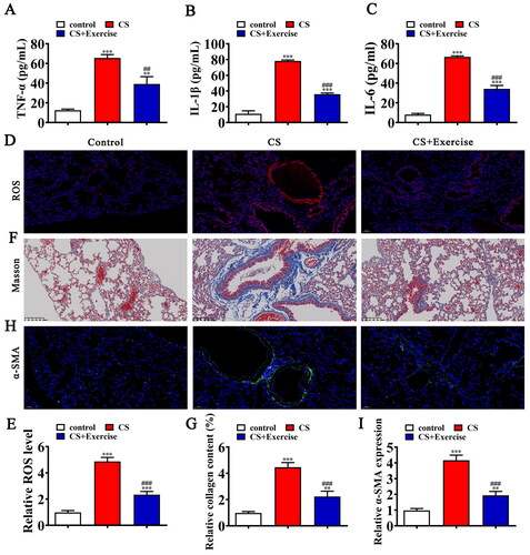
Accumulation study found that exercise can improves physical performance ability, shortness of breath, and the quality of life in patients with COPD [Citation9, Citation10]. In this study, we also found that inflammatory factor TNF-α, IL-1β, IL-6 expression were increased in cigarette smoking (CS) induced COPD mice with ELISA detection. And exercise significantly reversed COPD induced inflammatory factor expression (). Immunofluorescence for ROS detection shows that exercise significantly reversed COPD induced ROS level in lung tissues (). Masson trichrome staining showed that as CS exposure increased the small air ways of mice became thicker and collagen accumulated. Exercise significantly improves COPD induced pulmonary fibrosis (). Immunofluorescence assessment showed increased α-SMA expression in CS induced COPD mice, which suggested an increase in airway epithelial cells to mesenchymal transition. Exercise significantly reversed COPD induced α-SMA expression in lung tissues ().
Figure 1. Exercise ameliorates COPD induced lung injury by inhibit ROS, inflammation and epithelial-mesenchymal transition (EMT). (A–C) ELISA detection shows the expression of inflammatory factor TNF-α, IL-1β, IL-6. Data are means ± SD. **p < 0.01, ***p < 0.001 vs. control. ##p < 0.01, ###p < 0.001 vs. CS. (D and E) Immunofluorescence detection shows the ROS level in lung tissues. Data are means ± SD. ***p < 0.001 vs. control. ###p < 0.001 vs. CS. (F and G) Representative images of lung sections after Masson trichrome staining. Data are expressed as mean ± SD. ***p < 0.001 vs. control. ##p < 0.01, ###p < 0.001 vs. CS. (H and I) Representative images and quantification of the α-SMA expression in the lung tissue samples in different groups. Data are expressed as mean ± SD. **p < 0.01, ***p < 0.001 vs. control. ###p < 0.001 vs. CS.
Downregulation lnc-H19 inhibits COPD mediated lung injury
Previous study has found that lnc-H19 expression was increased in human pulmonary fibrotic tissues [Citation11, Citation12]. In this study we also found that lnc-H19 expression was increased in lung tissues in CS induced COPD mice. Exercise significantly reversed COPD induced lnc-H19 expression in lung tissues (). Then we constructed siRNA against lnc-H19 (si-lnc-H19) and transfected into BECs. RT-qPCR detection shows that lnc-H19 expression was increased in BECs after CSE solution treatment. After transfected with si-lnc-H19, the expression of lnc-H19 significantly decreased ().
Figure 2. Downregulation lnc-H19 inhibits COPD mediated lung injury. (A) RT-qPCR detection shows the expression of lnc-H19 among normal, CS, and CS + Exercise mice serum. Data are means ± SD. **p < 0.01 vs. control. ##p < 0.01 vs. CS. (B) RT-qPCR detection shows the expression of lnc-H19 in BECs. Data are means ± SD. ***p < 0.001 vs. NC. ###p < 0.001 vs. CSE. (C–E) ELISA detection shows the expression of inflammatory factor TNF-α, IL-1β, IL-6. Data are means ± SD. **p < 0.01, ***p < 0.001 vs. NC. ###p < 0.001 vs. CSE. (F and G) Immunofluorescence detection shows the ROS level in lung tissues. Data are means ± SD. ***p < 0.001 vs. NC. ###p < 0.001 vs. CSE. (H and I) Representative images and quantification of the α-SMA expression in the skin tissue samples in different groups. Data are means ± SD. **p < 0.01, ***p < 0.001 vs. NC. ###p < 0.001 vs. CSE.
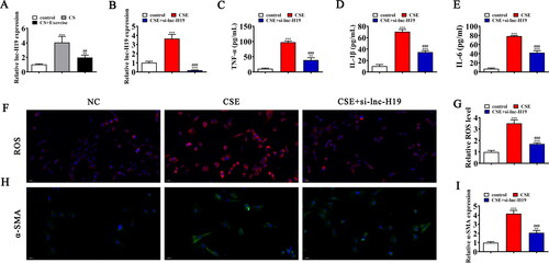
ELISA detection shows that inflammatory factor TNF-α, IL-1β, IL-6 expression were increased in CSE solution treated BECs. But lnc-H19 silence reversed CSE induced inflammatory factor expression (). Immunofluorescence for ROS detection shows that lnc-H19 silence reversed CSE induced ROS expression (). Immunofluorescence assessment also showed that lnc-H19 silence reversed CSE induced α-SMA expression in BECs ().
miR-181 and PDCD4 were downstream targets of lnc-H19
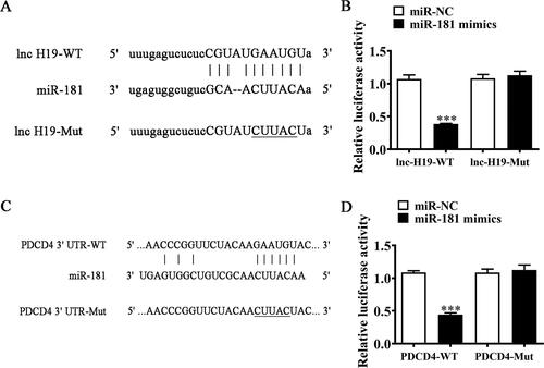
The accumulation study has been confirmed that lncRNAs act as miRNA sponge in order to modulate mRNA expression [Citation13]. In this study, bioinformatics analysis found that lnc-H19 can interacted with different miRNA including miR-181. Then we constructed luciferase reporter vector and the result shows that miR-181 can decreased luciferin activity (). Suggestion that miR-181 was the downregulation target of lnc-H19. Further study also found that miR-181 can interacted with 3′UTR of PDCD4. Then we constructed luciferase reporter vector and the result shows that miR-181 can decreased luciferin activity (). Suggestion that PDCD4 was the downregulation target of miR-181.
Figure 3. miR-181 and PDCD4 were downstream targets of lnc-H19. (A) Bioinformatics analysis predicting binding sites of miR-181 in lnc-H19. Mutant version of lnc-H19 is shown. (B) Relative luciferase activity determined 48 hours after transfection of BECs cells with miR-181 mimic/NC or lnc-H19 wild-type/Mut. Data are means ± SD. ***p < 0.01. (C) Prediction of miR-181 binding sites in the 3′-UTR of PDCD4. Mutant version of 3′-UTR-PDCD4 is shown. (D) Relative luciferase activity 48 hours after transfection of BECs with miR-181 mimic/NC or 3′-UTR-PDCD4 wild-type/Mut. Data are means ± SD. ***p < 0.01.
Upregulation of PDCD4 or downregulation of miR-181 reversed the protective effect of si-lnc-H19 to BECs after exposure to CSE
RT-qPCR results showed that lnc-H19 expression was decreased after transfected with si-lnc-H19. But treatment with the miR-670-5p inhibitor or PDCD4 overexpression vector (PDCD4) has no effect to lnc-H19 expression in BECs cells (). Suggestion that both miR-181 and PDCD4 were the downstream target of lnc-H19. Rt-qPCR detection also found that lnc-H19 silence increased miR-181 expression. PDCD4 upregulation cannot restore miR-181 expression after lnc-H19 silence (). Suggestion that miR-181 was at the downstream of lnc-H19. The result also found that lnc-H19 silence decreased PDCD4 expression. But inhibitor miR-181 restored PDCD4 expression after si-lnc-H19 (). Suggestion that lnc-H19 promotion PDCD4 expression by sponging miR-181.
Figure 4. Upregulation of PDCD4 or downregulation of miR-181 reversed the protective effect of si-lnc-H19 to BECs after exposure to CSE. (A–C) RT-qPCR detection shows the expression of lnc-H19, miR-181, and PDCD4 in BECs. Data are means ± SD. ***p < 0.001 vs. NC. ###p < 0.001 vs si-lnc-H19. (D-F) ELISA detection shows the expression of inflammatory factor TNF-α, IL-1β, IL-6. Data are means ± SD. **p < 0.01, ***p < 0.001 vs. NC. ##p < 0.01, ###p < 0.001 vs si-lnc-H19. (G and H) Immunofluorescence detection shows the ROS level in BECs. Data are means ± SD. *p < 0.05, ***p < 0.001 vs. NC. ###p < 0.001 vs si-lnc-H19. (I and J) Representative images and quantification of the α-SMA expression in the BECs in different groups. Data are means ± SD. *p < 0.05, ***p < 0.001 vs. NC. ###p < 0.001 vs si-lnc-H19.
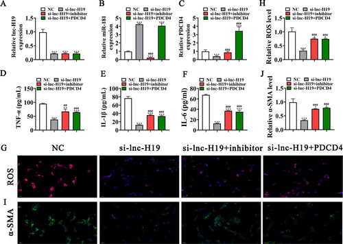
ELISA detection shows that inflammatory factor TNF-α, IL-1β, IL-6 expression were decreased in CSE pretreatment BECs cells after silence lnc-H19. Upregulation of PDCD4 or downregulation of miR-181 reversed the protective effect of si-lnc-H19 to inflammatory factor expression in BECs after exposure to CSE (). Immunofluorescence for ROS detection shows that upregulation of PDCD4 or downregulation of miR-181 restored CSE induced ROS expression after lnc-H19 silence (). Immunofluorescence assessment also showed that upregulation of PDCD4 or downregulation of miR-181 restored CSE induced α-SMA expression after lnc-H19 silence ().
Discussion
COPD is one of the leading causes of death worldwide [Citation14]. Oxidative stress (ROS) and inflammation imbalance leads to the pathogenesis of multiple pulmonary diseases such as asthma, chronic obstructive pulmonary disease (COPD) [Citation15, Citation16]. In this study we found that ROS and inflammatory factor TNF-α, IL-1β, IL-6 expression in lung tissues were increased in cigarette smoking (CS) induced COPD mice. Exercise ameliorates COPD induced lung injury by partial inhibition ROS and inflammatory factor expression. The study also found that exercise can reversed COPD induced extracellular matrix remodeling (epithelial-mesenchymal transition (EMT)) and pulmonary fibrosis. Previous studies have found that EMT act as a primary mechanism of COPD [Citation17, Citation18]. Downregulation of epithelial phenotypes (E-cadherin, ZO-1, and occludin), increased mesenchymal phenotypes (vimentin, α-smooth muscle actin [α-SMA], and fibronectin), and extracellular matrix remodeling, as well as altered barrier function, are all associated with CS-induced COPD phenotypes [Citation19].
Previous studies have found that lnc-H19 plays a role in microenvironmental regulation [Citation20]. This study found that lnc-H19 expression was increased in lung tissues of COPD mice. Exercise decreased COPD induced lnc-H19 expression in lung tissues of COPD mice. Accumulation study found that these lncRNA-miRNA-mRNA networks are potential biomarkers for microenvironmental regulation [Citation21–23]. In this study, Bioinformatics analysis and luciferase report analysis found that miR-181 and PDCD4 were downstream targets of lnc-H19. lnc-H19 downregulation promote miR-181 expression. Downregulation miR-181 restored the ROS, inflammatory cytokines and EMT relative protein α-SMA expression in si-lnc-H19 BECs after exposure to CSE. Previous studies have been confirmed that miR-181 expression was decreased in COPD patient and the expression of miR-181 can alleviating inflammatory diseases [Citation24, Citation25].
Further study has been found that PDCD4 was the downstream target of miR-181. lnc-H19 downregulation decreased PDCD4 expression. But inhibitor miR-181 restored PDCD4 expression after si-lnc-H19. Previous studies have found that PDCD4 expression suppressing Nrf2 and further promotion ROS, inflammatory cytokines, and EMT [Citation26–29]. In this study, we found that upregulation of PDCD4 or downregulation of miR-181 restored CSE induced ROS, inflammatory cytokines, and EMT relative protein α-SMA expression after lnc-H19 silence. Suggestion that lnc-H19 expression promotion COPD induced lung injury by inducing miR-181/PDCD4 axis.
Conclusion
In conclusion, lnc-H19 expression causes dysfunction in the lungs, as well as abnormal mesenchymal transition induced by subchronic CS exposure. Upregulation of PDCD4 or downregulation of miR-181 reversed the protective effect of si-lnc-H19 to BECs after exposure to CSE. We validated the therapeutic potential of lnc-H19 in a CS-induced COPD by regulation miR-181/PDCD4 signaling which increased ROS, inflammatory cytokines expression, and EMT.
Author’s contributions
P. L. and L. Z. contributed to the study conception and design. All authors collected the data and performed the data analysis. All authors contributed to the interpretation of the data and the completion of figures and tables. All authors contributed to the drafting of the article and final approval of the submitted version.
Ethics approval and consent to participate
Animal experimentation was approved by the Ethics Committee of the Shanghai Gongli Hospital.
Disclosure statement
All the authors declare that they have no conflict of interest.
Data availability
The datasets used and/or analyzed during the current study are available from the corresponding author on reasonable request.
Additional information
Funding
References
- Rabe KF, Watz H. Chronic obstructive pulmonary disease. Lancet. 2017;389(10082):1931–1940. DOI:10.1016/S0140-6736(17)31222-9
- Altawalbeh SM, Hijazi B, Kufoof L, et al. Health expenditures of asthma-COPD overlap in Northern Jordan. PLoS One. 2021;16(9):e0257566. DOI:10.1371/journal.pone.0257566
- Burke H, Wilkinson TMA. Unravelling the mechanisms driving multimorbidity in COPD to develop holistic approaches to patient-centred care. Eur Respir Rev. 2021;30(160):210041. DOI:10.1183/16000617.0041-2021
- Roman-Rodriguez M, Kaplan A. GOLD 2021 strategy report: implications for asthma-COPD overlap. Int J Chron Obstruct Pulmon Dis. 2021;16:1709–1715. DOI:10.2147/COPD.S300902
- Borghi-Silva A, Garcia-Araújo AS, Winkermann E, et al. Exercise-based rehabilitation delivery models in comorbid chronic pulmonary disease and chronic heart failure. Front Cardiovasc Med. 2021;8:729073. DOI:10.3389/fcvm.2021.729073
- Raveling T, Vonk J, Struik FM, et al. Chronic non-invasive ventilation for chronic obstructive pulmonary disease. Cochrane Database Syst Rev. 2021;8(8):Cd002878. DOI:10.1002/14651858.CD002878.pub3
- Watson A, Wilkinson TMA, Freeman A. Evidence around the impact of pulmonary rehabilitation and exercise on redox status in COPD: a systematic review. Front Sports Act Living. 2021;3:782590. DOI:10.3389/fspor.2021.782590
- Su W, Huo Q, Wu H, et al. The function of LncRNA-H19 in cardiac hypertrophy. Cell Biosci. 2021;11(1):153. DOI:10.1186/s13578-021-00668-4
- Chen S, Liu D, Zhou Z, et al. Role of long non-coding RNA H19 in the development of osteoporosis. Mol Med. 2021;27(1):122. DOI:10.1186/s10020-021-00386-0
- Adolfo JR, Dhein W, Sbruzzi G. Intensity of physical exercise and its effect on functional capacity in COPD: systematic review and meta-analysis. J Bras Pneumol. 2019;45(6):e20180011. DOI:10.1590/1806-3713/e20180011
- Poulet C, Njock MS, Moermans C, et al. Exosomal long non-coding RNAs in lung diseases. Int J Mol Sci. 2020;21(10):3580. DOI:10.3390/ijms21103580
- Wang X, Cheng Z, Dai L, et al. Knockdown of long noncoding RNA H19 represses the progress of pulmonary fibrosis through the transforming growth factor β/Smad3 pathway by regulating MicroRNA 140. Mol Cell Biol. 2019;39(12):e00143–19. DOI:10.1128/MCB.00143-19
- Huang Y. The novel regulatory role of lncRNA-miRNA-mRNA axis in cardiovascular diseases. J Cell Mol Med. 2018;22(12):5768–5775. DOI:10.1111/jcmm.13866
- Cassady SJ, Reed RM. Pulmonary hypertension in COPD: a case study and review of the literature. Medicina (Kaunas, Lithuania). 2019;55(8):432. DOI:10.3390/medicina55080432
- Zuo L, Wijegunawardana D. Redox role of ROS and inflammation in pulmonary diseases. Adv Exp Med Biol. 2021;1304:187–204. DOI:10.1007/978-3-030-68748-9_11
- Tian X, Xue Y, Xie G, et al. (-)-epicatechin ameliorates cigarette smoke-induced lung inflammation via inhibiting ROS/NLRP3 inflammasome pathway in rats with COPD. Toxicol Appl Pharmacol. 2021;429:115674. DOI:10.1016/j.taap.2021.115674
- Hou W, Hu S, Li C, et al. Cigarette smoke induced lung barrier dysfunction, EMT, and tissue remodeling: a possible link between COPD and lung cancer. Biomed Res Int. 2019;2019:2025636. DOI:10.1155/2019/2025636
- Shukla S, Ward C, Walters EH. Mechanistic insights on EMT and smoking-related COPD. Stem Cell Rev Rep. 2021;17(4):1503–1504. DOI:10.1007/s12015-021-10152-8
- Wang Q, Sundar IK, Lucas JH, et al. Molecular clock REV-ERBα regulates cigarette smoke-induced pulmonary inflammation and epithelial-mesenchymal transition. JCI Insight. 2021;6(12):e145200. DOI:10.1172/jci.insight.145200
- Lu J, Wang Y, Hu Y, et al. Lnc-H19 enhances anaerobic glycolysis of keloid fibroblasts by targeting the miR-214-5p/FGF2 axis. Burns. 2021. DOI:10.1016/j.burns.2021.07.015
- Ma N, Tie C, Yu B, et al. Identifying lncRNA-miRNA-mRNA networks to investigate Alzheimer’s disease pathogenesis and therapy strategy. Aging (Albany NY). 2020;12(3):2897–2920. 72920. DOI:10.18632/aging.102785
- Wang JY, Yang Y, Ma Y, et al. Potential regulatory role of lncRNA-miRNA-mRNA axis in osteosarcoma. Biomed Pharmacother. 2020;121:109627. DOI:10.1016/j.biopha.2019.109627
- Zheng Y, Zhang Y, Zhang X, et al. Novel lncRNA-miRNA-mRNA competing endogenous RNA triple networks associated programmed cell death in heart failure. Front Cardiovasc Med. 2021;8:747449. DOI:10.3389/fcvm.2021.747449
- Ma XF, Qin J, Guo XH. MiR-181-5p protects mice from sepsis via repressing HMGB1 in an experimental model. Eur Rev Med Pharmacol Sci. 2020;24(18):9712–9720.
- Zhao S, Lin C, Yang T, et al. Expression of long non-coding RNA LUCAT1 in patients with chronic obstructive pulmonary disease and its potential functions in regulating cigarette smoke extract-induced 16HBE cell proliferation and apoptosis. J Clin Lab Anal. 2021;35(7):e23823. DOI:10.1002/jcla.23823
- Hwang SK, Jeong YJ, Chang YC. PDCD4 inhibits lung tumorigenesis by the suppressing p62-Nrf2 signaling pathway and upregulating Keap1 expression. Am J Cancer Res. 2020;10(2):424–439.
- Cho JH, Kim YW, Choi BY, et al. Sulforaphane inhibition of TPA-mediated PDCD4 downregulation contributes to suppression of c-Jun and induction of p21-dependent Nrf2 expression. Eur J Pharmacol. 2014;741:247–253. DOI:10.1016/j.ejphar.2014.08.007
- Jain S, Durugkar S, Saha P, et al. Effects of intranasal azithromycin on features of cigarette smoke-induced lung inflammation. Eur J Pharmacol. 2022;915:174467. DOI:10.1016/j.ejphar.2021.174467
- Yeo CD, Kim JW, Ha JH, et al. Chemopreventive effect of phosphodieasterase-4 inhibition in benzo(a)pyrene-induced murine lung cancer model. Exp Lung Res. 2014;40(10):500–506. DOI:10.3109/01902148.2014.950769