Abstract
The etiology of sex differences in the risk of asthma-COPD phenotype and COPD is still not completely understood. Genetic and environmental risk factors are commonly believed to play an important role. This study aims to identify sex-specific genetic markers associated with asthma-COPD phenotype and COPD using the Canadian Longitudinal Study on Aging (CLSA) Baseline Comprehensive and Genomic data. There were a total of 1,415 COPD cases. Out of them, 504 asthma-COPD phenotype cases were identified. 20,524 participants without a diagnosis of asthma and COPD served as controls. We performed genome-wide SNP-by-sex interaction analysis. SNPs with an interaction p-value < 10−5 were included in a sex-stratified multivariable logistic regression for asthma-COPD phenotype and COPD outcomes. 18 and 28 SNPs had a significant interaction term p-value < 10−5 with sex in the regression analyses of asthma-COPD phenotype and COPD outcomes, respectively. Sex-stratified multivariable analysis of asthma-COPD phenotype showed that 7 SNPs in/near SMYD3, FHIT, ZNF608, RIMBP2, ZNF133, BPIFB1, and S100B loci were significant in males. Sex-stratified multivariable analysis of COPD showed that 8 SNPs in/near MAGI1, COX18, OSTC, ELOVL5, C7orf72 FGF14, and NKAIN4 were significant in males, and 4 SNPs in/near genes CAMTA1, SATB2, PDE10A, and LINC00908 were significant in females. An SNP in the ZPBP gene was associated with COPD in both males and females. Identification of sex-specific loci associated with asthma-COPD phenotype and COPD may offer valuable evidence toward a better understanding of the sex-specific differences in the pathophysiology of the diseases.
Introduction
Chronic obstructive pulmonary disease (COPD) is a chronic respiratory condition characterized by irreversible airflow limitation. It is a prevalent disease among middle-aged and elderly. In 2015, GINA and GOLD collaboration described a new chronic obstructive airway phenotype known as asthma-COPD overlap (Citation1). The Asthma-COPD phenotype was characterized as a chronic obstructive airway phenotype with persistent airflow limitation and a clinical presentation of features associated with asthma and COPD. A number of studies have shown that patients with the overlap phenotype present with more frequent airflow obstruction symptoms, more exacerbation, lower quality of life, recurrent hospitalization, and more medical utilization than patients with classic COPD or asthma alone (Citation2–7). In addition, the lack of clinical data from randomized controlled trials explicitly addressing treatment regimens for patients with the overlap phenotype makes definitive treatment difficult.
Many epidemiological studies have found significant sex differences in the risk of asthma-COPD phenotype and COPD, with females having a higher prevalence than males (Citation8–12). COPD was initially considered a disease of middle-aged and older men; however, its prevalence and hospitalization rates in women have increased in the last 20 years (Citation13–15). Although the etiology of sex differences is not fully understood, it is commonly believed that both environmental and genetic factors play an important role.
Cigarette smoking is the most consistent risk factor for COPD, especially in developed nations; exposure to biomass and noxious pollutants impacts COPD risk in developing countries (Citation16,Citation17). Apart from the strong influence of environmental factors on COPD risk, various genetic risk factors have been associated with COPD susceptibility (Citation18–22). As genetic and environmental risk factors play crucial roles in COPD development, studies have revealed that COPD’s clinical presentation and susceptibility vary by gender (Citation23–31). Women who were ex-smokers and current smokers had a higher risk of airflow obstruction than men who were ex-smokers and current smokers exposed to the same dose of tobacco (Citation31). A study observed that females with early-onset COPD and < 20 pack-years of smoking had a greater reduction in lung function (FEV1% predicted) and more severe disease than males (Citation24). Females with COPD have been reported to have more dyspnea, less expectoration, worse exercise capacity, and more frequent diagnosis of concomitant asthma than males (Citation29). Whereas in another study, males with COPD were reported to have more emphysema and lesser airway remodeling than females (Citation28). Epidemiological studies have linked several socio-demographic, socio-economic, and lifestyle factors to asthma-COPD phenotype risk (Citation8,Citation9). In a study of the risk of asthma-COPD phenotype in Aboriginal people, daily smoking, working more than 40 h per week, and being separated, widowed, or divorced were associated with the risk of asthma-COPD phenotype in females but not in males (Citation9). In addition, Aboriginal women who smoked at home compared to those who did not were almost three times more likely to be associated with asthma-COPD phenotype risk. However, this trend was not observed in men (Citation9). Similarly, in another population-based study, women who smoked cigarettes for ten years or more had a higher prevalence of asthma-COPD phenotype than men (Citation12). Nevertheless, the underlying mechanisms for the sex-specific differences in asthma-COPD phenotype and COPD remain unclear.
Previous research has identified various genetic loci associated with COPD and asthma-COPD phenotype (Citation18–22,Citation32–34). However, no genetic association studies have examined sex-specific genetic risk factors for asthma-COPD phenotype while including socio-demographic, socio-economic, and lifestyle factors in their analyses. Using the Canadian Longitudinal Study on Aging (CLSA) baseline questionnaire and genomic dataset, this study aimed to explore sex-specific genetic differences in asthma-COPD phenotype and COPD while accounting for various socio-demographic, lifestyle, socio-economic, and environmental factors.
Materials and method
Study design and population
The Canadian Longitudinal Study on Aging (CLSA) is a population-based cohort of 51,338 Canadians aged 45 to 85 years old. The CLSA is made up of two complementary cohorts: the Tracking cohort, which consisted of 21,241 people who were questioned over the phone, and the Comprehensive cohort, which consisted of 30,097 people who provided baseline data via an in-person home interview as well as other questionnaires, tests, physical measurements, and biospecimen (blood and urine) collected at the data collection sites (Citation35). Our study focused on a subset of 26,622 subjects from the Comprehensive Cohort who were genotyped using the Affymetrix UK Biobank Axiom array (Citation36).
Outcomes
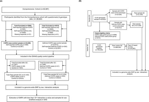
We defined COPD and asthma from the positive responses to the questions, “Has a doctor told you that you have/had any of the following: emphysema, chronic bronchitis, chronic obstructive pulmonary disease (COPD), or chronic changes in the lungs due to smoking?” and “Has a doctor ever told you had asthma?”, respectively. Participants with positive responses to both self-reported physician-diagnosed asthma and COPD were categorized as asthma-COPD phenotype. Control subjects were those who responded “no” to self-reported physician diagnoses of asthma and COPD. Participants who self-reported a physician diagnosis of only asthma and those with missing responses were excluded from each study group. Asthma-COPD phenotype cases were also excluded from the COPD case-control group. On the other hand, COPD cases were excluded from the asthma-COPD phenotype case-control group (see ).
Predictor variables
The CLSA questionnaire contains a wide range of socio-demographic and socio-economic data, information on lifestyle and health behaviors, as well as clinical and physical data. Our study considered the following potential confounders such as age group (45 to 54 years, 55 to 64 years, 65 to 74 years, and 75 years and above), biological sex, marital status (single or never married, married or in a common-law relationship, widowed/divorced/separated), an education level (less than post-secondary education, post-secondary but not university education, and university education/others), total personal income and total household income (Less than $20,000, $20,000 to less than $50,000, $50,000 to less than $100,000, $100,000 or more), province of recruitment (Prairies, British Columbia, Eastern provinces, Ontario and Quebec), retirement status (Retired completely and not retired/partly retired), smoking status (current, never, and former smokers), homeownership (owned and rented/others), and urban/rural dwelling.
Genotyping and quality control
Genotyping for 794,409 genetic markers was done using Affymetrix UK Biobank Axiom array. Quality control of genetic markers and samples was adequately described in another paper (Citation36). SNPs and Samples that failed the QC metrics were removed. All genomic positions were in reference to the human genome build GRCh37/hg19. Using PLINK v1.90b6.25 (Citation37), we excluded samples with discordant sex information and more than 5% genotype missingness. Autosomal SNPs were extracted, SNPs with less than 99% call rate, minor allele frequency less than 1% (MAF < 0.01), and SNPs deviating from the Hardy Weinberg equilibrium threshold of 1e-10 were excluded. In order to obtain SNPs that are in approximate linkage equilibrium with each other, we generated a subset of LD pruned SNPs using PLINK’s Indep-pairwise command (Indep-pairwise 50, 5, 0.5). Following quality control, we had 504 asthma-COPD phenotype cases, 911 COPD cases, 20,524 controls, and 416,562 SNPs for genome-wide SNP-by-sex interaction analysis (see ).
Genetic and statistical analysis
In order to investigate sex-specific variants for asthma-COPD phenotype and COPD outcomes, genome-wide SNP-by-Sex interaction was examined in logistic regression for the asthma-COPD phenotype and COPD, respectively, after adjusting for age, sex, smoking status, and the first four principal components of genetic ancestry using PLINK v1.90b6.25 (Citation37). Those SNPs with an interaction p value less than 10−5 were further examined in a sex-stratified analysis for asthma-COPD phenotype and COPD outcomes using survey-specific logistic regression (Proc Surveylogistic) in SAS version 9.4. The survey-specific SAS procedure allows us to apply sampling weights, incorporate complex survey designs, and control for potential confounders. For SNPs in high linkage disequilibrium (D’>0.80), the SNP with the highest polymorphism information content (PIC) value was chosen.
For descriptive statistics of continuous and categorical variables, mean and standard error and frequency and percentage were used to describe the study population. CLSA’s trimmed inflation and analytic weights (CLSA Sample Weights Version 1.2) were used for descriptive and regression analysis. In order to identify potential confounders to be included in the final sex-stratified analysis, a univariate logistic regression model was used to identify significant risk factors. The predictor variables with a p value ≤ 0.20 from the univariate analysis were entered into an interim multivariate model. The least significant variable was then removed one at a time until only variables with significant p value (p ≤ 0.05) and clinically important factors remained in the model.
In order to discover genetic risk factors that are specific to either sex, a survey logistic regression was conducted with sex as a domain factor. For the SNPs with an interaction term p value less than 10−5, we included one SNP at a time and the first four principal components into the final model while controlling for potential environmental, demographic, and socio-economic risk factors. In addition to the four principal components, the final model for the asthma-COPD phenotype included adjustments for age, smoking status, marital status, homeownership, total household income, education level, and retirement status. For the COPD outcome, the variables adjusted for were age, smoking status, province of recruitment, marital status, homeownership, total personal income, and retirement status.
Three inheritance models (dominant, recessive, and additive) for each variant were considered. The model with the lowest Akaike information criterion values (AIC) was regarded as the best-fitting model. The results from the best-fitted model were presented.
In order to account for multiple comparisons, the Bonferroni correction was applied in the sex-stratified analyses of asthma-COPD phenotype and COPD based on the number of SNPs with a significant interaction term with the sex variable. We created regional association plots using Locuszoom (Citation38) for the significant SNPs from the sex-stratified analyses. The strength of association was reported as odds ratios with 95% confidence intervals.
Results
Characteristics of the study population
depicts the characteristics of the study population stratified by sex. A total of 26622 subjects (13,343 males and 13,279 females) represented 3,295,958 participants in the weighted sample (1,670,060 males and 1,625,898 females).
Table 1. Descriptive characteristics of the study population.
In males and females, the prevalence of asthma-COPD phenotype was 1.4% and 2.3%, respectively. The prevalence of COPD, however, was 2.6% for males and 3.0% for females.
Males and females differed significantly in the distribution of age, age groups, smoking status, marital status, urban or rural dwelling, homeownership, total household income, total personal income, retirement status, province of recruitment, and highest education status.
Approximately 52.4% of males were smokers (current smokers: 10% and former smokers:42.4%), whereas, in females, 48.6% had a positive smoking history (current smokers: 8.5% and former smokers: 40.1%). In comparison to males, there were more females in the age groups of 55 to 64 (30.2% vs. 29.8%), 65 to 74 (18.3% vs. 15.8%), and 75 and over (11.7% vs. 10.2%). In contrast, more males (44.3%) than females (39.7%) were in the age group 45-54. A significantly higher proportion of females (21.6%) were widowed/divorced/separated than males (9.6%).
Males were more likely than females to have a total household income (49.1% vs. 38.0%) and a total personal income (23.6% vs. 8.2%) of $100,000 or more. In contrast, more females than males earned less than $20,000 (5.6% vs. 3.4%) and less than $50,000 (22.2% vs. 15.1%) for total household income. Similarly, for total personal income, a higher proportion of females than males earned less than $20,000 (22.9% vs. 7.8%) and less than $50,000 (39.9% vs. 27.9%). Females (38.0%) tended to be retired more than males (29.0%). In contrast, for the highest education status, males (61.4%) were more likely to have post-secondary education at the university level than females (56.8%).
Genome-wide SNP-by-sex interaction
In the SNP-by-sex interaction analysis, we examined after quality control, 416,562 SNPs, 21435 subjects for COPD (cases: n = 911, controls: n = 20524) and 21028 subjects for the asthma-COPD phenotype (cases: n = 504, controls: n = 20524) (see ).
There were 18 and 28 distinct SNPs from the SNP-by-sex interaction terms with p value less than 10¯5 for asthma-COPD phenotype and COPD, respectively (see ). No indication of population stratification based on the genomic inflation factor was observed in the genome-wide interaction analysis for both phenotypes (see ).
Figure 2. Quantile-Quantile plots (Q-Q plot) showing the distribution of observed versus Expected pvalue. (A) Q-Q plot for the genome-wide SNP-by-sex interaction for asthma-COPD phenotype. Genomic inflation factor, lambda (λ) = 1.00. (B) Q-Q plot for the genome-wide SNP-by-sex interaction for COPD. Genomic inflation factor, lambda (λ) = 1.00.
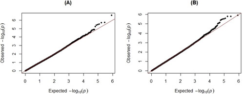
Table 2. 18 Signals of the SNP-by-Sex Interactions with pvalue less than 10¯5 for asthma-COPD phenotype.
Table 3. 28 Signals of the SNP-by-Sex Interactions with pvalue less than 10−5 for COPD.
SNP rs926718, an intronic variant in the ZNF133 gene, had the lowest interaction p value (p = 2.18 × 10¯5) for the asthma-COPD phenotype (). Regarding COPD outcome, the SNP with the lowest p value was an intronic variant [rs73838466 (p = 9.35 × 10¯6)] in the OSTC gene ().
Sex stratified analysis for asthma-COPD phenotype
Out of the 18 SNPs with a significant SNP-by-sex interaction term, pvalue less than 10¯5 for asthma-COPD phenotype, the polymorphisms rs61140467, which was in high LD (D’=0.99) with rs11799559, was excluded from the analysis due to its lower polymorphic information content. demonstrates the results of significant SNPs from the sex-stratified multivariate analysis of asthma-COPD phenotype after Bonferroni correction (p ≤ 0.003 = 0.05/17). 7 SNPs were significant in males, and no SNP was significant in females (see and Supplementary Tables S3 and S4). The regional plots of these 7 male-specific SNPs associated with asthma-COPD phenotype are shown in .
Table 4. Result of sex-stratified analysis for asthma-COPD phenotype.
The top significant SNP amongst the males was rs3821479 in the FHIT gene [OR = 0.55, p < 0.0001, 95% CI (0.41 − 0.73)]. The polymorphisms rs926718 in the ZNF133 gene, which had the lowest SNP-by-sex interaction pvalue, was significantly associated with asthma-COPD phenotype amongst the males [OR = 0.58, p = 0.0005, 95% CI (0.43 − 0.79)]. Howbeit, the effects of these variants were protective ( and ).
Figure 3. Forest plot showing the direction of effects of the male-specific SNPs associated with asthma-COPD phenotype from the sex-stratified analysis.
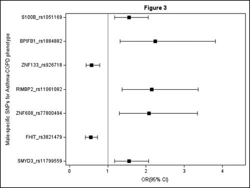
As illustrated in , SNP rs11061082 in/near the RIMBP2 gene [OR = 2.15, p = 0.0009, 95% CI (1.37 − 3.37)] and SNP rs1884882 near BPIFB1 gene [OR = 2.24, p = 0.0029, 95% CI (1.32 − 3.81)] under the recessive inheritance model were significantly associated with asthma-COPD phenotype risk in males. While in the additive model, SNPs rs11799559 [OR = 1.56, p = 0.0021, 95% CI (1.18 − 2.07)] in the SMYD3 gene, rs1051169 [OR = 1.56, p = 0.0019, 95% CI (1.18 − 2.06)] in S100B gene and rs77800494 [OR = 2.08, p = 0.0025, 95% CI (1.30 − 3.34)] near ZNF608 gene were significantly associated with asthma-COPD phenotype risk in males (see ).
Sex-stratified analysis for COPD
Out of the 28 SNPs with a significant SNP-by-sex interaction term, pvalue less than 10¯5 for COPD, the polymorphism rs73838466, which was in high LD (D’=0.99) with rs17039240, was excluded from the analysis due to its lower polymorphic information content. displays the results of significant SNPs from the sex-stratified multivariate analysis of COPD after Bonferroni correction (p ≤ 0.002 = 0.05/27).
Table 5. Result of sex-stratified analysis for COPD.
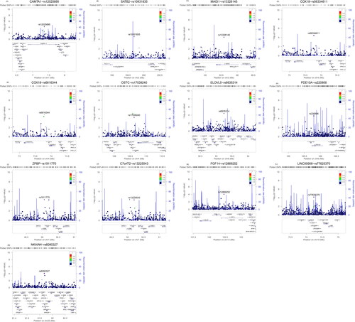
As illustrated in , and , eight variants (rs13326145, rs56334611, rs6816344, rs17039240, rs6935314, rs13225543, rs12869252, and rs6090327) mapped in/near MAGI1, COX18, OSTC, ELOVL5, C7orf72 FGF14, and NKAIN4 genes respectively were significantly associated with COPD in males. Whereas four SNPs (rs12025895, rs10931835, rs220806, and rs77625370) in/near CAMTA1, SATB2, PDE10A, and LINC00908 genes were significantly associated with COPD amongst females. presents a regional plot of significant sex-specific SNPs associated with COPD and their genomic positions relative to genes or nearby genes. The SNP rs1911770 in gene ZPBP was found to have a statistically significant association with COPD in males [OR = 1.64, p = 0.0005, 95% CI (1.24 − 2.16)] and females [OR = 0.76, p = 0.002, 95% CI (0.64 − 0.91)]; indicating that it increased the risk of COPD in males while conferring a protective effect in females (see ).
Figure 4. Forest plots showing the direction of effects for the sex-specific SNPs associated with COPD from the sex-stratified analysis. (A) Males. (B) Females. Only significant SNPs after the Bonferroni adjustment were plotted.
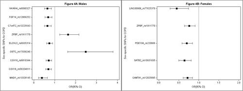
Figure 5. Regional plots for the 7 male-specific SNPs associated with asthma-COPD phenotype showing their respective genomic locations. (A) rs11799559 in SMYD3 gene. (B) rs3821479 in FHIT gene. (C) rs77800494 near ZNF608 gene. (D) rs11061082 near RIMBP2 gene. (E) rs926718 in ZNF133 gene. (F) rs1884882 near BPIFB1 gene. (G) rs1051169 in S100B gene.
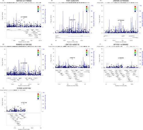
Figure 6. Regional plots for the sex-specific SNPs associated with COPD showing their respective genomic locations (A) rs12025895 in CAMTA1 gene. (B) rs10931835 near SATB2 gene. (C) rs13326145 in MAGI1 gene. (D) rs56334611 near COX18 gene. (E) rs6816344 near COX18 gene. (F) rs17039240 near OSTC gene. (G) rs6935314 near ELOVL5 gene. (H) rs220806 in PDE10A gene. (I) rs1911770 in ZPBP gene. (J) rs13225543 near C7orf72 gene. (K) rs12869252 in FGF14 gene. (L) rs77625370 near LINC00908 gene. (M) rs6090327 in NKAIN4 gene.
Out of the eight male-specific SNPs, an intergenic SNP rs17039240 near the OSTC gene was significantly associated with COPD risk in males [OR= 2.48, p < 0.0001, 95% CI (1.62 − 3.79)] but not in females (see ). The top female-specific SNP was rs12025895 in the CAMTA1 gene [OR = 0.72, p < 0.0001, 95% CI (0.61 − 0.84)]. All the variants significantly associated with COPD amongst females had protective effects, with ORs ranging from 0.45 to 0.76 (see ).
Discussion
This study found 18 and 28 distinct signals for a genome-wide SNP-by-Sex interaction on asthma-COPD phenotype and COPD outcomes, respectively. The SNPs with the lowest SNP-by-Sex interaction pvalue for asthma-COPD phenotype and COPD outcomes were located in the intronic regions of the ZNF133 and OTSC genes at 20p11.23 and 4q25 cytogenetic positions, respectively.
We discovered seven male-specific variants in or adjacent SMYD3, FHIT, ZNF608, RIMBP2, ZNF133, BPIFB1, and S100B genes that were significantly associated with asthma-COPD phenotype. Five of the seven SNPs (rs11799559, rs77800494, rs11061082, rs1884882, and rs1051169) were associated with an increased risk, with ORs ranging from 1.56 to 2.24, while the remaining two SNPs (rs3821479 and rs926718) showed protective effects, with OR of 0.55 and 0.58.
A total of eight male-specific variants (rs13326145, rs56334611, rs6816344, rs17039240, rs6935314, rs13225543, rs12869252, and rs6090327) in/near the MAGI1, COX18, OSTC, ELOVL5, C7orf72 FGF14, and NKAIN4 genes, and 4 female-specific variants (rs12025895, rs10931835, rs220806 and rs77625370) in/near the CAMTA1, SATB2, PDE10A, and LINC00908 genes were significantly associated with COPD. In addition, rs1911770 in the ZPBP gene, was associated with an increased risk of COPD in males and a reduced risk in females.
To the best of our knowledge, this is the first study to look at sex-specific genetic risk factors for asthma-COPD phenotype and COPD while also taking into account environmental, lifestyle, socio-economic, and socio-demographic factors. Previous genetic and gene-based association studies had demonstrated sex-specific genetic effects on COPD and asthma (Citation39–41); however, these studies did not consider the influence of socio-demographic and socio-economic factors.
In this study, the strongest male-specific associations for asthma-COPD phenotype risk were from SNPs (rs1884882, rs11061082 rs77800494, rs11799559, rs1051169) in/near BPIFB1, RIMBP2, ZNF608, SMYD3, and S100B genes. The BPIFB1 gene on chromosome 20q11.21 encodes a protein secreted by goblet cells in the airway epithelium, trachea, submucosal glands of airways, and nasal cavities (Citation42). This protein is believed to play a role in innate immunity against inhaled toxins and pathogens. BPIFBI is upregulated in several respiratory diseases. For example, after a segmental allergen challenge, higher levels of BPIFB1 were found in bronchoalveolar lavage fluid in asthmatic patients (Citation43). In addition, BPIFB1 levels in the sputum of smokers with COPD were significantly higher than in smokers and nonsmokers without COPD (Citation44). De Smet et al. (Citation45) observed that the mRNA expression levels of BFIFB1 amongst COPD subjects were positively correlated with disease severity and that smokers with COPD had higher BPIFB1 mRNA and protein expression levels in lung tissue and airway epithelium than nonsmokers and smokers without COPD. BPIFB1 levels have also been found to be significantly inversely correlated to FEV1% predicted, FEV1/FVC ratio, and diffusing capacity of the lung for carbon monoxide (DLCO), all of which are proxies for COPD disease severity and emphysema (Citation44,Citation45). Additionally, human and animal studies have demonstrated that males are more prone to emphysematous changes than females (Citation28,Citation46).
Polymorphisms of RIMBP2, FHIT, and ZNF608 genes on 12q24.33, 3p14.2, and 5q23.2 cytogenetic positions have been associated with testosterone levels (Citation47). Several studies have suggested the influence of sex hormones on lung diseases and inflammatory responses of the lungs to pathogens and inhaled toxins, including cigarette smoke and pollutants (Citation48,Citation49). Androgen (testosterone) is thought to have anti-inflammatory effects, which are mediated by interaction with androgen receptors (AR) and control the expression of transcriptions (Citation50). For example, one study found that testosterone reduced pulmonary epithelial inflammation in rats with COPD (Citation51). As androgens decline with advancing age, the binding of androgen and AR complexes to transcriptions might be attenuated. These might lead to altered expressions of genes, increased pro-inflammatory cytokines, and chronic inflammatory diseases.
The S100B gene on chromosome 21q22.3 belongs to the S100 family of proteins, which regulates calcium balance, cell apoptosis, migration, proliferation, differentiation, energy metabolism, and inflammation. Furthermore, research has indicated that S100B is a major ligand of the receptor for advanced glycation end products (RAGE), a pattern recognition receptor that is expressed in alveolar type I and type II epithelial cells, bronchiolar epithelium, and alveolar macrophages (Citation52,Citation53). S100 protein interaction with RAGE activates NF-κB, causing the production of pro-inflammatory cytokines and the migration of neutrophils, monocytes, and macrophages (Citation54). For instance, In an in-vitro study, S100B was shown to stimulate the secretion of TNF-alpha and IL-6 in Alveolar Type-I (AT-I) derived cells from the pulmonary tissue of male fetuses of Han-Wistar rats (Citation55). The SMYD3 gene, a protein-coding gene known for its histone methyltransferase activity, is more expressed in males’ dorsolateral frontal cortex and anterior cingulate cortex of the brain than in females (Citation56). Other histone-encoding genes have been shown to be more expressed in males than females in the heart, kidney, and colon (Citation56).
In this study, the strongest male-specific SNP for COPD was rs17039240. This SNP is an intergenic variant near the OSTC gene. OSTC plays a critical role in the generation and processing of amyloid-beta peptides (Aβ) from the amyloid precursor protein (APP) (Citation57). APP and Aβ have not been adequately explored in lung diseases; however, studies have shown that APP and Aβ from human monocyte-derived macrophages regulate pro-inflammatory and anti-inflammatory mediators (Citation58). Increased levels of amyloid-beta peptides have been observed in the serum and lungs of COPD patients compared to controls. In addition, higher serum Aβ negatively correlated with worse lung function in COPD patients (Citation59,Citation60). Studies have shown that androgens regulate amyloid beta-peptides (Citation61–63). Gillett et al. (Citation61) demonstrated that lower androgen levels were associated with increased plasma amyloid beta-peptide in older men with dementia. Furthermore, low total testosterone has been associated with worse pulmonary function in men with COPD (Citation64). Given that a significant proportion of COPD patients are middle-aged and older men, the age-related decline in androgens (low testosterone) in males, as well as the proteolytic action of OSTC protein on amyloid precursor protein, may result in elevated levels of amyloid beta-peptide, suggesting a potential role of OSTC gene polymorphisms in increasing COPD risk in males.
We also observed intronic and intergenic SNPs (rs1911770 and rs13225543) in/near ZPBP and C7orf72 genes associated with COPD in males. ZPBP, a Zona Pellucida Binding Protein implicated in adult fertility, is expressed in the testis and ovary (Citation65,Citation66). This intronic SNP (rs1911770) in the ZPBP gene had the opposite effect on the risk of COPD in males and females (increasing COPD risk for males but protective for females). In a previous GWAS, variants in/near the ZPBP gene approached genome-wide significance for an association with pulmonary function amongst smokers (FEV1 and FEV1/FVC ratio) (Citation67). In addition, ZPBP is a paralog to ZPBP2, which is located on chromosome 17q21.1. Research has shown that ZPBP2 is associated with asthma and childhood asthma (Citation68,Citation69). In a study conducted by Naumova et al. (Citation70), they discovered sex-specific differences in the DNA methylation of the ZPBP2 gene in relation to asthma susceptibility. Specifically, males were revealed to have a lower average methylation than females in the ZPBP2 gene promoter region, implying that hypo-methylation of the ZPBP2 gene increases asthma risk in males. Furthermore, ZPBP/ZPBP2 deletion in a mouse model produces sperm abnormalities and infertility in men but not females (Citation65).
The intergenic SNP (rs13225543) near the C7orf72 gene was found to be male-specific. C7orf72 gene, a spermatogenesis-associated protein, has been linked to spermatogenesis (Citation71). Studies indicate that in male hamsters, pulmonary emphysema affects spermatogenesis, resulting in morphophysiological changes to the reproductive organs due to increased oxidative stress and testosterone imbalance (Citation72). This suggests that the polymorphisms of ZPBP and C7orf72 may have a stronger impact on the development of diseases in males than in females.
Other male-specific associations, albeit protective in the direction of effect, were discovered within or near the MAGI1, COX18, ELOVL5, FGF14, and NKAIN4 genes in the 3p14.1, 4q13.3, 6p12.1, 13q33.1, and 20q13.33 cytogenetic bands, respectively. Variants of the FGF14 gene, which belongs to the fibroblast growth factor family, have been associated with post-BD FEV1 in children with asthma (Citation73). Members of the FGF family have been associated with lung development and respiratory disease (Citation74,Citation75). For instance, polymorphisms in the FGF7 gene have been reported to be associated with COPD (Citation74). FGFs 1,2,8,9 and 10 have been implicated in various levels of lung development (Citation75). Interestingly, FGF10 expression was higher in males than females in a study examining the expression profile of androgen-regulated genes in murine fetal developing lungs (Citation76). In our study, an SNP (rs12869252) in FGF14 was significantly associated with COPD in males and may, in combination with sex hormones, potentially play a sexually dimorphic role in COPD susceptibility.
MAGI1 is widely expressed in lung epithelial cells, where it functions as a scaffolding protein at intercellular junctions and maintains epithelial barrier function (Citation77). The airway epithelial lining serves as the first line of defense against environmental insults such as cigarette smoke. MAGI1 gene has been implicated as a surfactant regulator with increased expression in the fetal lung of males compared to females (Citation76). Cigarette smoke, a significant risk factor for COPD, adversely affects surfactants and airway epithelial architecture (Citation78–80). In one study, the expression of the MAGI1 gene in the airway epithelium was significantly downregulated in smokers with COPD and healthy smokers compared to nonsmokers (Citation80). This suggests that cigarette smoke compromises the integrity of airway epithelial cell-cell junction. With males and females having differential susceptibility to cigarette smoke, the distribution and population of MAGI1 proteins in airway epithelial cells’ tight junctions may play an important role in COPD pathogenesis in males and females.
Two SNPs, rs56334611 and rs6816344, located near the COX18 gene, were associated with COPD in males. COX18 gene encodes a cytochrome c oxidase assembly protein responsible for mitochondrial biogenesis, MT-CO2/COX2 maturation, and regulation of mitochondrial respiratory chain complex IV. Oxidative stress due to excessive reactive oxygen species in COPD patients has been linked to mitochondrial damage, reduced mitochondrial biogenesis, and mitochondrial homeostasis (Citation81,Citation82). Sex disparities in oxidative stress and reactive oxygen species generation have been reported, with males having more oxidative stress, more reactive oxygen species, and lower antioxidant capacity than females (Citation83). ELOVL5 gene, widely expressed in the brain, lung, testis, adrenal gland, and prostate, also regulates mitochondrial functions and reactive oxygen species production (Citation84,Citation85). ELOVL5 has also been found to be overexpressed in prostate cancer. For instance, one experimental study discovered that ELOVL5 was significantly more expressed in prostate cancer cells than in normal/benign prostatic hyperplasia cells and that this upregulation was mediated via androgen receptors (Citation84).
Variants (rs12025895, rs10931835, rs220806 and rs77625370) within/near the CAMTA1 SATB2 PDE10A and LINC00908 genes on chromosomes 1p36.31, 2q33.1, 6q27, and 18q23, respectively, showed female-specific associations with COPD. In a large GWAS of lung function using the UK biobank data, SNPs in SATB2 have been associated with an increase in FEV1 and FVC (Citation32). In an experimental study using ovariectomized rats, Wu et al. (Citation86) demonstrated that the bone marrow stromal cells (BMSCs) of ovariectomized rats experienced weaker SATB2 expression, reduced bone formation capacity, and increased senescence. On the other hand, estrogen increased SATB2 expression, slowed down cellular aging, and increased the osteogenicity of bone marrow stromal cells. Estrogen deficiency has been associated with osteoporosis during post-menopause (Citation87). Osteoporosis is a major comorbid condition in females with COPD (Citation88). It is likely that the expression of the SATB2 gene may decrease as estrogen levels decrease in menopausal and post-menopausal females with COPD, potentially resulting in a decline in lung function.
CAMTA1 gene, another female-specific association with COPD in our study, has been associated with lung function and COPD (Citation89,Citation90). Kang et al. (Citation91) suggested that the CAMTA1 gene plays a regulatory role in the nuclear factor of activated T cells pathway. The nuclear factor of activated T cells (NFAT), identified in activated T-cells, regulates the expression of IL-2, IL-4, and IL-5 (Citation92,Citation93). T-cells play a central role in an adaptive immune response. Also, Innate and adaptive immune responses differ between sexes. Females have been reported to have more activated T-cells and T-cell proliferation than males (Citation94). Studies have shown that the increased number of T cells in the lungs and airways of patients with COPD correlates with disease severity (Citation95). Furthermore, it has been found that female smokers appear to experience a higher level of inflammatory responses in the airways than their male counterparts (Citation27). These suggest that the CAMTA1 gene may modulate inflammatory mechanisms differently for males and females with COPD.
An SNP (rs220806) in the PDE10A gene was one of the female-specific loci for COPD. This gene’s protein belongs to the cyclic nucleotide phosphodiesterase family (PDEs), which plays an important role in controlling intracellular cyclic nucleotide by hydrolyzing cAMP and cGMP second messengers involved in regulating airway smooth muscle function (Citation96). PDE10A plays an essential role in lung inflammation by promoting macrophage activation and neutrophil infiltration (Citation97). For example, PDE10A knockout mice exhibited reduced IL-1b, MCP-1, IL-6, and TNF-alpha protein levels in lung tissues than in PDE010-WT mice after exposure to lipopolysaccharide (Citation97). Sexual dimorphism in the PDE10A gene has been demonstrated in an experimental animal study. PDE10A knockout mice were confirmed to have decreased body weight compared to their wild-type counterparts, with females being more affected than males (Citation98).
In our study, the identified sex-specific loci associated with asthma-COPD phenotype and COPD may have direct or indirect sexually dimorphic roles. This suggests that the complex interplay between these sex-specific gene signatures and sex hormones or lifestyle factors, such as cigarette smoking, may influence the varying expression and pathobiological functions of these genes in males and females, thus leading to differences in susceptibility to asthma-COPD phenotype and COPD.
Our study had some limitations. The identification of COPD and asthma-COPD phenotype cases as self-reported physician-diagnosed COPD and concomitant diagnosis of COPD and asthma without objective spirometry measurements are subject to misclassification. In this case, a non-differential misclassification (i.e., equal chance of misclassification of our outcome between those with polymorphisms and those with wild-type), might result in reduced statistical power and an underestimation of the observed association. However, large GWAS and population-based studies have widely used self-reported obstructive airway disease diagnosed by physicians to identify genetic and clinical characteristics (Citation11,Citation12,Citation99–102). Self-reported asthma has been shown to have high reliability and validity (Citation103). In addition, self-reported physician-diagnosed COPD/emphysema/chronic bronchitis has been demonstrated to have very high specificity and low sensitivity (Citation104). A recent population study of individuals aged 50–64 years concluded that self-reported physician-diagnosed COPD is a valid tool in studies of risk factors for COPD in the general population, not in studies of prevalence of COPD, due to its high specificity (>95%) and low sensitivity (<13%) (Citation104). This implies that false positives will be reduced. However, the low sensitivity will cause the prevalence of COPD to be underestimated. Nonetheless, our primary goal was to identify sex-specific genetic risk factors.
The sample size for our study was moderately large. However, it appeared relatively underpowered to identify variants (rare or common) with interaction p value significant at the genome-wide significance threshold. Genome-wide interaction studies, in general, necessitate a much larger sample size and more statistical power than standard GWAS. Due to the high proportion of missing values in the pre-bronchodilator spirometry variables and the absence of post-bronchodilator spirometry parameters in the dataset, we could not use spirometry criteria to define our outcomes. Our study lacked an independent replication cohort to validate our findings. Future replication of our findings in subsequent studies could improve generalizability.
Conclusion
Our study identified novel sex-specific loci associated with asthma-COPD phenotype and COPD. These findings are potential precursors to deepening our understanding of sex-related genetic differences in asthma-COPD phenotype and COPD pathology. Future research exploring the expression quantitative trait loci (eQTLs) and discovering the functional sex-specific roles of these genetic signatures may improve disease endotyping in individuals with asthma-COPD phenotype and COPD.
Ethical approval
This CLSA project received ethics approval at two levels. Consent to participate was obtained for all participants under the CLSA harmonized multi-university ethics process approved by the Hamilton Integrated Research Ethics Board (HiREB), Hamilton Health Sciences/McMaster University. Simon Fraser University (SFU) was a participating institution in the CLSA data collection, and the SFU Office of Research Services Ethics Committee reviewed all consent material prior to data collection (SFU ORS #2018s0139). Ethics approval for the current study was obtained from the University of Alberta Health Research Ethics Board (Pro00091377_REN3) and Memorial University Health Research Ethics Board (HREB # 2019.072).
Disclaimer
The opinions expressed in this manuscript are the author’s own and do not reflect the views of the Canadian Longitudinal Study on Aging.
Supplemental Material
Download PDF (506.2 KB)Acknowledgment
This research was made possible using the data/biospecimens collected by the Canadian Longitudinal Study on Aging (CLSA). This research has been conducted using CLSA’s Baseline Comprehensive Dataset version 4.0 and Genomics Dataset version 3.0, under Application Number 19CA006. The CLSA is led by Drs. Parminder Raina, Christina Wolfson, and Susan Kirkland. The opinions expressed in this manuscript are solely those of the author and do not represent those of the Canadian Longitudinal Study on Aging.
Disclosure statement
The authors declare no conflicts of interest.
Data availability statement
The datasets used in this study are not readily available. Data are only available from the Canadian Longitudinal Study on Aging (www.clsa-elcv.ca) for researchers who meet the criteria for access to de-identified CLSA data.
Additional information
Funding
References
- GOLD_ACOS. _2015.pdf, https://goldcopd.org/wp-content/uploads/2016/04/GOLD_ACOS_2015.pdf. (accessed 17 May 2022).
- Hardin M, Silverman EK, Barr RG, et al. The clinical features of the overlap between COPD and asthma. Respir Res. 2011;12(1):8. doi: 10.1186/1465-9921-12-127.
- Papaiwannou A, Zarogoulidis P, Porpodis K, et al. Asthma-chronic obstructive pulmonary disease overlap syndrome (ACOS): current literature review. J Thorac Dis. 2014;6: s 146–S151.
- Rhee CK, Yoon HK, Yoo KH, et al. Medical utilization and cost in patients with overlap syndrome of chronic obstructive pulmonary disease and asthma. COPD. 2014;11(2):163–170. doi: 10.3109/15412555.2013.831061.
- Bujarski S, Parulekar AD, Sharafkhaneh A, et al. The asthma COPD overlap syndrome (ACOS). Curr Allergy Asthma Rep. 2015;15(3):7. doi: 10.1007/s11882-014-0509-6.
- de Marco R, Marcon A, Rossi A, et al. Asthma, COPD and overlap syndrome: a longitudinal study in young European adults. Eur Respir J. 2015;46(3):671–679. doi: 10.1183/09031936.00008615.
- Lange P, Çolak Y, Ingebrigtsen TS, et al. Long-term prognosis of asthma, chronic obstructive pulmonary disease, and asthma-chronic obstructive pulmonary disease overlap in the Copenhagen city heart study: a prospective population-based analysis. Lancet Respir Med. 2016;4(6):454–462. doi: 10.1016/S2213-2600(16)00098-9.
- Koleade A, Farrell J, Mugford G, et al. Prevalence and risk factors of ACO (Asthma-COPD overlap) in aboriginal people. J Environ Public Health. 2018;2018:4657420. doi: 10.1155/2018/4657420.
- Koleade A, Farrell J, Mugford G, et al. Female-specific risk factors associated with risk of ACO (asthma COPD overlap) in aboriginal people. J Asthma. 2020;57(9):925–932. doi: 10.1080/02770903.2019.1621890.
- de Marco R, Pesce G, Marcon A, et al. The coexistence of asthma and chronic obstructive pulmonary disease (COPD): prevalence and risk factors in young, middle-aged and elderly people from the general population. PLOS One. 2013;8(5):e62985. doi: 10.1371/journal.pone.0062985.
- Senthilselvan A, Beach J. Characteristics of asthma and COPD overlap syndrome (ACOS) in the Canadian population. J Asthma. 2019;56(11):1129–1137. doi: 10.1080/02770903.2018.1531997.
- Wheaton AG, Pleasants RA, Croft JB, et al. Gender and asthma-chronic obstructive pulmonary disease overlap syndrome. J Asthma. 2016;53(7):720–731. doi: 10.3109/02770903.2016.1154072.
- Aryal S, Diaz-Guzman E, Mannino DM. Influence of sex on chronic obstructive pulmonary disease risk and treatment outcomes. Int J Chron Obstruct Pulmon Dis. 2014;9:1145–1154. doi: 10.2147/COPD.S54476.
- Ntritsos G, Franek J, Belbasis L, et al. Gender-specific estimates of COPD prevalence: a systematic review and meta-analysis. Int J Chron Obstruct Pulmon Dis. 2018;13:1507–1514. doi: 10.2147/COPD.S146390.
- Ford ES, Croft JB, Mannino DM, et al. COPD surveillance—United States, 1999-2011. Chest. 2013;144(1):284–305. doi: 10.1378/chest.13-0809.
- DeMeo DL. Sex and gender omic biomarkers in men and women with COPD. Chest. 2021;160(1):104–113. doi: 10.1016/j.chest.2021.03.024.
- Salvi SS, Barnes PJ. Chronic obstructive pulmonary disease in non-smokers. Lancet. 2009;374:11.
- Silverman EK. Genetics of COPD. Annu Rev Physiol. 2020;82:413–431. doi: 10.1146/annurev-physiol-021317-121224.
- Hall R, Hall IP, Sayers I. Genetic risk factors for the development of pulmonary disease identified by genome-wide association. Respirology. 2019;24(3):204–214. doi: 10.1111/resp.13436.
- Sakornsakolpat P, Prokopenko D, Lamontagne M, et al. Genetic landscape of chronic obstructive pulmonary disease identifies heterogeneous cell-type and phenotype associations. Nat Genet. 2019;51(3):494–505. doi: 10.1038/s41588-018-0342-2.
- Cho MH, McDonald MLN, Zhou X, et al. Risk loci for chronic obstructive pulmonary disease: a genome-wide association study and meta-analysis. Lancet Respir Med. 2014;2(3):214–225. doi: 10.1016/S2213-2600(14)70002-5.
- Odimba U, Senthilselvan A, Farrell J, et al. Current knowledge of Asthma-COPD overlap (ACO) genetic risk factors, characteristics, and prognosis. COPD. 2021;18(5):585–595. doi: 10.1080/15412555.2021.1980870.
- Aryal S, Diaz-Guzman E, Mannino DM. COPD and gender differences: an update. Transl Res. 2013;162(4):208–218. doi: 10.1016/j.trsl.2013.04.003.
- Sørheim IC, Johannessen A, Gulsvik A, et al. Gender differences in COPD: are women more susceptible to smoking effects than men? Thorax. 2010;65(6):480–485. doi: 10.1136/thx.2009.122002.
- Silverman EK, Weiss ST, Drazen JM, et al. Gender-related differences in severe, early-onset chronic obstructive pulmonary disease. Am J Respir Crit Care Med. 2000;162(6):2152–2158. doi: 10.1164/ajrccm.162.6.2003112.
- Hardin M, Foreman M, Dransfield MT, et al. Sex-specific features of emphysema among current and former smokers with COPD. Eur Respir J. 2016;47(1):104–112. doi: 10.1183/13993003.00996-2015.
- Gut-Gobert C, Cavaillès A, Dixmier A, et al. Women and COPD: do we need more evidence? Eur Respir Rev. 2019;28(151):180055. doi: 10.1183/16000617.0055-2018.
- Martinez FJ, Curtis JL, Sciurba F, et al. Sex differences in severe pulmonary emphysema. Am J Respir Crit Care Med. 2007;176(3):243–252. doi: 10.1164/rccm.200606-828OC.
- Perez TA, Castillo EG, Ancochea J, et al. Sex differences between women and men with COPD: a new analysis of the 3CIA study. Respir Med. 2020;171:106105. doi: 10.1016/j.rmed.2020.106105.
- Gan WQ, Man SP, Postma DS, et al. Female smokers beyond the perimenopausal period are at increased risk of chronic obstructive pulmonary disease: a systematic review and meta-analysis. Respir Res. 2006;7(1):9. doi: 10.1186/1465-9921-7-52.
- Amaral AFS, Strachan DP, Burney PGJ, et al. Female smokers are at greater risk of airflow obstruction than male smokers. UK Biobank. Am J Respir Crit Care Med. 2017;195(9):1226–1235. doi: 10.1164/rccm.201608-1545OC.
- Shrine N, Guyatt AL, Erzurumluoglu AM, et al. New genetic signals for lung function highlight pathways and chronic obstructive pulmonary disease associations across multiple ancestries. Nat Genet. 2019;51(3):481–493. doi: 10.1038/s41588-018-0321-7.
- Hardin M, Cho M, McDonald ML, et al. The clinical and genetic features of COPD-asthma overlap syndrome. Eur Respir J. 2014;44(2):341–350. doi: 10.1183/09031936.00216013.
- John C, Guyatt AL, Shrine N, et al. Genetic associations and architecture of Asthma-COPD overlap. Chest. 2022;161(5):1155–1166. doi: 10.1016/j.chest.2021.12.674.
- Raina P, Wolfson C, Kirkland S, et al. Cohort profile: the Canadian longitudinal study on aging (CLSA). Int J Epidemiol. 2019;48(6):1752–1753j. doi: 10.1093/ije/dyz173.
- Forgetta V, Li R, Darmond-Zwaig C, et al. Cohort profile: genomic data for 26 622 individuals from the Canadian longitudinal study on aging (CLSA). BMJ Open. 2022;12(3):e059021. doi: 10.1136/bmjopen-2021-059021.
- Purcell S, Chang C. General usage - PLINK 1.9, https://www.cog-genomics.org/plink/1.9/general_usage. (accessed 23 May 2022).
- Pruim RJ, Welch RP, Sanna S, et al. LocusZoom: regional visualization of genome-wide association scan results. Bioinformatics. 2010;26(18):2336–2337. doi: 10.1093/bioinformatics/btq419.
- Joo J, Himes B. Gene-based analysis reveals sex-specific genetic risk factors of COPD. AMIA Annu Symp Proc. 2021;2021:601–610.
- Hardin M, Cho MH, Sharma S, et al. Sex-Based genetic association study identifies CELSR1 as a possible chronic obstructive pulmonary disease risk locus among women. Am J Respir Cell Mol Biol. 2017;56(3):332–341. doi: 10.1165/rcmb.2016-0172OC.
- Myers RA, Scott NM, Gauderman WJ, et al. Genome-wide interaction studies reveal sex-specific asthma risk alleles. Hum Mol Genet. 2014;23(19):5251–5259. doi: 10.1093/hmg/ddu222.
- Bingle CD, Wilson K, Lunn H, et al. Human LPLUNC1 is a secreted product of goblet cells and minor glands of the respiratory and upper aerodigestive tracts. Histochem Cell Biol. 2010;133(5):505–515. doi: 10.1007/s00418-010-0683-0.
- Wu J, Kobayashi M, Sousa EA, et al. Differential proteomic analysis of bronchoalveolar lavage fluid in asthmatics following segmental antigen challenge*S. Mol Cell Proteomics. 2005;4(9):1251–1264. doi: 10.1074/mcp.M500041-MCP200.
- Gao J, Ohlmeier S, Nieminen P, et al. Elevated sputum BPIFB1 levels in smokers with chronic obstructive pulmonary disease: a longitudinal study. Am J Physiol Lung Cell Mol Physiol. 2015;309(1):L17–L26. doi: 10.1152/ajplung.00082.2015.
- De Smet EG, Seys LJ, Verhamme FM, et al. Association of innate defense proteins BPIFA1 and BPIFB1 with disease severity in COPD. Int J Chron Obstruct Pulmon Dis. 2018;13:11–27. doi: 10.2147/COPD.S144136.
- Tam A, Churg A, Wright JL, et al. Sex differences in airway remodeling in a mouse model of chronic obstructive pulmonary disease. Am J Respir Crit Care Med. 2016;193(8):825–834. doi: 10.1164/rccm.201503-0487OC.
- Ruth KS, Day FR, Tyrrell J, et al. Using human genetics to understand the disease impacts of testosterone in men and women. Nat Med. 2020;26(2):252–258. doi: 10.1038/s41591-020-0751-5.
- Fuentes N, Silveyra P. Endocrine regulation of lung disease and inflammation. Exp Biol Med. 2018;243(17-18):1313–1322. doi: 10.1177/1535370218816653.
- Becerra-Diaz M, Song M, Heller N. Androgen and androgen receptors as regulators of monocyte and macrophage biology in the healthy and diseased lung. Front Immunol. 2020;11:1698. doi: 10.3389/fimmu.2020.01698.
- Morooka N, Ueguri K, Yee KKL, et al. Androgen-androgen receptor system improves chronic inflammatory conditions by suppressing monocyte chemoattractant protein-1 gene expression in adipocytes via transcriptional regulation. Biochem Biophys Res Commun. 2016;477(4):895–901. doi: 10.1016/j.bbrc.2016.06.155.
- Wang X, Huang L, Jiang S, et al. Testosterone attenuates pulmonary epithelial inflammation in male rats of COPD model through preventing NRF1-derived NF-κB signaling. J Mol Cell Biol. 2021;13(2):128–140. doi: 10.1093/jmcb/mjaa079.
- Guo WA, Knight PR, Raghavendran K. The receptor for advanced glycation end products and acute lung injury/acute respiratory distress syndrome. Intensive Care Med. 2012;38(10):1588–1598. doi: 10.1007/s00134-012-2624-y.
- Leclerc E, Fritz G, Vetter SW, et al. Binding of S100 proteins to RAGE: an update. Biochim Biophys Acta. 2009;1793(6):993–1007. doi: 10.1016/j.bbamcr.2008.11.016.
- Xia C, Braunstein Z, Toomey AC, et al. S100 proteins as an important regulator of macrophage inflammation. Front Immunol. 2017;8:1908. doi: 10.3389/fimmu.2017.01908.
- Piazza O, Leggiero E, De Benedictis G, et al. S100B induces the release of pro-inflammatory cytokines in alveolar type I-like cells. Int J Immunopathol Pharmacol. 2013;26(2):383–391. doi: 10.1177/039463201302600211.
- Mayne BT, Bianco-Miotto T, Buckberry S, et al. Large scale gene expression Meta-analysis reveals tissue-specific, sex-biased gene expression in humans. Front Genet. 2016;7:183. doi: 10.3389/fgene.2016.00183.
- Wilson CM, Magnaudeix A, Yardin C, et al. DC2 and keratinocyte-associated protein 2 (KCP2), subunits of the oligosaccharyltransferase complex, are regulators of the γ-Secretase-directed processing of amyloid precursor protein (APP) *. J Biol Chem. 2011;286(36):31080–31091. doi: 10.1074/jbc.M111.249748.
- Spitzer P, Walter M, Göth C, et al. Pharmacological inhibition of amyloidogenic APP processing and knock-down of APP in primary human macrophages impairs the secretion of cytokines. Front Immunol. 2020;11:1967. doi: 10.3389/fimmu.2020.01967.
- Bu XL, Cao GQ, Shen LL, et al. Serum amyloid-beta levels are increased in patients with chronic obstructive pulmonary disease. Neurotox Res. 2015;28(4):346–351. doi: 10.1007/s12640-015-9552-x.
- Karoor V, Wick MJ, Sullivan T, et al. Cigarette smoke increases the expression of amyloid precursor protein in pulmonary artery smooth muscle cells. Am J Respir Crit Care Med. 2022;205: a 3021–A3021. Available from https://www.atsjournals.org/doi/pdf/10 .1164/ajrccm-conference.2022.205.1_MeetingAbstracts.A3021 doi: 10.1164/ajrccm-conference.2022.205.1_MeetingAbstracts.A3021.
- Gillett MJ, Martins RN, Clarnette RM, et al. Relationship between testosterone, sex hormone binding globulin and plasma amyloid beta peptide 40 in older men with subjective memory loss or dementia. J Alzheimers Dis. 2003;5(4):267–269. doi: 10.3233/jad-2003-5401.
- Ramsden M, Nyborg AC, Murphy MP, et al. Androgens modulate β-amyloid levels in male rat brain. J Neurochem. 2003;87(4):1052–1055. doi: 10.1046/j.1471-4159.2003.02114.x.
- Rosario ER, Pike CJ. Androgen regulation of β-amyloid protein and the risk of Alzheimer’s disease. Brain Res Rev. 2008;57(2):444–453. doi: 10.1016/j.brainresrev.2007.04.012.
- Atlantis E, Fahey P, Cochrane B, et al. Endogenous testosterone level and testosterone supplementation therapy in chronic obstructive pulmonary disease (COPD): a systematic review and meta-analysis. BMJ Open. 2013;3(8):e003127. doi: 10.1136/bmjopen-2013-003127.
- Lin YN, Roy A, Yan W, et al. Loss of zona pellucida binding proteins in the acrosomal matrix disrupts acrosome biogenesis and sperm morphogenesis. Mol Cell Biol. 2007;27(19):6794–6805. doi: 10.1128/MCB.01029-07.
- Fowler PA, Flannigan S, Mathers A, et al. Gene expression analysis of human fetal ovarian primordial follicle formation. J Clin Endocrinol Metab. 2009;94(4):1427–1435. doi: 10.1210/jc.2008-2619.
- Lutz SM, Cho MH, Young K, et al. A genome-wide association study identifies risk loci for spirometric measures among smokers of European and African ancestry. BMC Genet. 2015;16(1):11. doi: 10.1186/s12863-015-0299-4.
- Verlaan DJ, Berlivet S, Hunninghake GM, et al. Allele-specific chromatin remodeling in the ZPBP2/GSDMB/ORMDL3 locus associated with the risk of asthma and autoimmune disease. Am J Hum Genet. 2009;85(3):377–393. doi: 10.1016/j.ajhg.2009.08.007.
- Yan Q, Brehm J, Pino-Yanes M, et al. A meta-analysis of genome-wide association studies of asthma in puerto ricans. Eur Respir J. 2017;49(5):1601505. doi: 10.1183/13993003.01505-2016.
- Naumova AK, Al Tuwaijri A, Morin A, et al. Sex- and age-dependent DNA methylation at the 17q12-q21 locus associated with childhood asthma. Hum Genet. 2013;132(7):811–822. doi: 10.1007/s00439-013-1298-z.
- Zhang J, Yan R, Wu C, et al. Spermatogenesis-associated 48 is essential for spermatogenesis in mice. Andrologia. 2018;50(6):e13027. doi: 10.1111/and.13027.
- Vieira HR, Gonçalves GD, Vieira NA, et al. Pulmonary emphysema impairs male reproductive physiology due to testosterone and oxidative stress imbalance in mesocricetus auratus. Reprod Sci. 2020;27(11):2052–2062. doi: 10.1007/s43032-020-00224-4.
- Brehm JM, Man Tse S, Croteau-Chonka DC, et al. A genome-wide association study of post-bronchodilator lung function in children with asthma. Am J Respir Crit Care Med. 2015;192(5):634–637. doi: 10.1164/rccm.201501-0047LE.
- Brehm JM, Hagiwara K, Tesfaigzi Y, et al. Identification of FGF7 as a novel susceptibility locus for chronic obstructive pulmonary disease. Thorax. 2011;66(12):1085–1090. doi: 10.1136/thoraxjnl-2011-200017.
- Yang L, Zhou F, Zheng D, et al. FGF/FGFR signaling: from lung development to respiratory diseases. Cytokine Growth Factor Rev. 2021;62:94–104. doi: 10.1016/j.cytogfr.2021.09.002.
- Bresson E, Seaborn T, Côté M, et al. Gene expression profile of androgen modulated genes in the murine fetal developing lung. Reprod Biol Endocrinol. 2010;8:2. doi: 10.1186/1477-7827-8-2.
- Laura RP, Ross S, Koeppen H, et al. MAGI-1: a widely expressed, alternatively spliced tight junction protein. Exp Cell Res. 2002;275(2):155–170. doi: 10.1006/excr.2002.5475.
- Moré JM, Voelker DR, Silveira LJ, et al. Smoking reduces surfactant protein D and phospholipids in patients with and without chronic obstructive pulmonary disease. BMC Pulm Med. 2010;10:53. doi: 10.1186/1471-2466-10-53.
- Bowler RP. Surfactant protein D as a biomarker for chronic obstructive pulmonary disease. COPD. 2012;9(6):651–653. doi: 10.3109/15412555.2012.736317.
- Shaykhiev R, Otaki F, Bonsu P, et al. Cigarette smoking reprograms apical junctional complex molecular architecture in the human airway epithelium in vivo. Cell Mol Life Sci. 2011;68(5):877–892. doi: 10.1007/s00018-010-0500-x.
- Barnes PJ. Oxidative stress in chronic obstructive pulmonary disease. Antioxidants. 2022;11(5):965. doi: 10.3390/antiox11050965.
- Jiang Y, Wang X, Hu D. Mitochondrial alterations during oxidative stress in chronic obstructive pulmonary disease. Int J Chron Obstruct Pulmon Dis. 2017;12:1153–1162. doi: 10.2147/COPD.S130168.
- Kander MC, Cui Y, Liu Z. Gender difference in oxidative stress: a new look at the mechanisms for cardiovascular diseases. J Cell Mol Med. 2017;21(5):1024–1032. doi: 10.1111/jcmm.13038.
- Centenera MM, Scott JS, Machiels J, et al. ELOVL5 is a critical and targetable fatty acid elongase in prostate cancer. Cancer Res. 2021;81(7):1704–1718. doi: 10.1158/0008-5472.CAN-20-2511.
- Di Gregorio E, Borroni B, Giorgio E, et al. ELOVL5 mutations cause spinocerebellar ataxia 38. Am J Hum Genet. 2014;95(2):209–217. doi: 10.1016/j.ajhg.2014.07.001.
- Wu G, Xu R, Zhang P, et al. Estrogen regulates stemness and senescence of bone marrow stromal cells to prevent osteoporosis via ERβ-SATB2 pathway. J Cell Physiol. 2018;233(5):4194–4204. doi: 10.1002/jcp.26233.
- Riggs BL. The mechanisms of estrogen regulation of bone resorption. J Clin Invest. 2000;106(10):1203–1204. doi: 10.1172/JCI11468.
- Raherison-Semjen C, Mezzi K, Kostikas K, et al. The perception of physicians on gender-specific differences in the diagnosis of COPD: results from a questionnaire-based survey. Int J Chron Obstruct Pulmon Dis. 2021;16:901–907. doi: 10.2147/COPD.S271505.
- Morrow JD, Glass K, Cho MH, et al. Human lung DNA methylation quantitative trait loci colocalize with chronic obstructive pulmonary disease genome-wide association loci. Am J Respir Crit Care Med. 2018;197(10):1275–1284. doi: 10.1164/rccm.201707-1434OC.
- Wang T, Wang W, Li W, et al. Genome-wide DNA methylation analysis of pulmonary function in middle and old-aged chinese monozygotic twins. Respir Res. 2021;22(1):300. doi: 10.1186/s12931-021-01896-5.
- Kang K, Peng X, Zhang X, et al. MicroRNA-124 suppresses the transactivation of nuclear factor of activated T cells by targeting multiple genes and inhibits the proliferation of pulmonary artery smooth muscle cells. J Biol Chem. 2013;288(35):25414–25427. doi: 10.1074/jbc.M113.460287.
- Barnes PJ. Transcription factors in airway diseases. Lab Invest. 2006;86(9):867–872. doi: 10.1038/labinvest.3700456.
- Lee JU, Kim LK, Choi JM. Revisiting the concept of targeting NFAT to control T cell immunity and autoimmune diseases. Front Immunol. 2018;9:2747. doi: 10.3389/fimmu.2018.02747.
- Klein SL, Flanagan KL. Sex differences in immune responses. Nat Rev Immunol. 2016;16(10):626–638. doi: 10.1038/nri.2016.90.
- Barnes PJ. Inflammatory mechanisms in patients with chronic obstructive pulmonary disease. J Allergy Clin Immunol. 2016;138(1):16–27. doi: 10.1016/j.jaci.2016.05.011.
- Billington CK, Ojo OO, Penn RB, et al. cAMP regulation of airway smooth muscle function. Pulm Pharmacol Ther. 2013;26(1):112–120. doi: 10.1016/j.pupt.2012.05.007.
- Hsu CG, Fazal F, Rahman A, et al. Phosphodiesterase 10A is a key mediator of lung inflammation. J Immunol. 2021;206(12):3010–3020. doi: 10.4049/jimmunol.2001026.
- Siuciak JA, McCarthy SA, Chapin DS, et al. Genetic deletion of the striatum-enriched phosphodiesterase PDE10A: evidence for altered striatal function. Neuropharmacology. 2006;51(2):374–385. doi: 10.1016/j.neuropharm.2006.01.012.
- Demenais F, Margaritte-Jeannin P, Barnes KC, et al. Multiancestry association study identifies new asthma risk loci that colocalize with immune-cell enhancer marks. Nat Genet. 2018;50(1):42–53. doi: 10.1038/s41588-017-0014-7.
- Moffatt MF, Gut IG, Demenais F, et al. A large-scale, consortium-based genomewide association study of asthma. N Engl J Med. 2010;363(13):1211–1221. doi: 10.1056/NEJMoa0906312.
- Hayden LP, Cho MH, Raby BA, et al. Childhood asthma is associated with COPD and known asthma variants in COPDGene: a genome-wide association study. Respir Res. 2018;19(1):209. doi: 10.1186/s12931-018-0890-0.
- Veerasingam E, Gao Z, Beach J, et al. Sex-specific characteristics for the coexistence of asthma and COPD in the Canadian population: a cross-sectional analysis of CLSA data. J Asthma. 2022;0:1–22.
- Mirabelli MC, Beavers SF, Flanders WD, et al. Reliability in reporting asthma history and age at asthma onset. J Asthma. 2014;51(9):956–963. doi: 10.3109/02770903.2014.930480.
- Torén K, Murgia N, Olin AC, et al. Validity of physician-diagnosed COPD in relation to spirometric definitions of COPD in a general population aged 50– 64 years - the SCAPIS pilot study. Int J Chron Obstruct Pulmon Dis. 2017;12:2269–2275. doi: 10.2147/COPD.S136308.


