To the Editor,
Primary germ cell tumours (GCTs) are rare malignant neoplasms occurring predominantly in children and adolescents. GCTs appear in the midline of the body, i.e. the gonads, retroperitoneum, mediastinum or the suprasellar and pineal region of the brain. Intracranial germ cell tumours are most often localised in the suprasellar region and may cause pituitary hormone deficits. They arise as a solitary nodule or less commonly as multiple lesions. They are thought to originate from a primordial germ cell that has mismigrated during embryonic development and later undergo malignant transformation. Some GCTs are capable of producing and secreting the tumour markers β-human chorionic gonadotropin (β-hCG) and/or α-fetoprotein (AFP). Elevated levels of tumour markers in serum and/or spinal fluid indicate the presence of a GCT [Citation1,Citation2].
Case report
In July 2007 a 19-year-old man was admitted to the hospital after a convulsive fit. The patient had a history of increasing headache for five months and daily vomiting for two months. Slight hydrocephalus was found on a computed tomography (CT)-scan. No neurological or visual deficit was detected. Magnetic resonance imaging (MRI) revealed multifocal lesions. A central tumour mass with a diameter of 34 mm located at the pineal region, and a large lesion partially situated intrasellarly and partially suprasellarly plus some smaller lesions related to the right lateral ventricle and the fourth ventricle were found. Aqueductal compression exerted by the pineal lesion caused obstructive hydrocephalus. The spinal canal showed no signs of pathology on MRI and no malignant cells were detected in the spinal fluid. The histopathological investigation of an endoscopic biopsy revealed germinoma. But because an increased level of AFP was found in the blood, the tumour was classified as a non-germinomatous germ cell tumour (NGGCT).
The endocrine abnormalities found initially were diabetes insipidus, a reduced level of thyroid hormones (T3 and T4) and a considerably increased level of prolactine. Later on, laboratory results revealed affection of most endocrine parameters. Synacten test was normal, but the gonadotropin and IGF-1 level were far below normal and the testosterone level in the blood was immeasurable. The patient had endocrine dysfunction during the entire disease course, and he was treated with hormone replacement therapy. No growth hormone substitution was given though, as long as the patient was in treatment of malignant disease. Four chemotherapy courses of cisplatin, etoposide and bleomycin (PEB) were administered with subsequent cerebral radiotherapy of 36 Gy. Additional boosts to the tumour areas of 18 Gy was given of 54 Gy in total.
Initially, β-hCG and AFP were within normal limits in the cerebrospinal fluid (CSF), while an increased concentration of AFP was detected in the serum. Based on these findings, an extracranial germ cell tumour secreting AFP was initially suspected. A CT-scan of thorax and abdomen along with ultrasound scan of the testicles, were carried out. The CT-scan was normal. Bilateral biopsy of the testes was performed and histopathological investigation revealed carcinoma in situ (CIS) in the left testicle and hypospermatogenesis in the right testicle. Shortly after end of chemotherapy, orchidectomy of the left testicle was performed and CIS testis without invasive growth was found in nearly all tubules.
In September 2007 an MRI of the brain showed significant regression of the tumours. AFP level in serum was normalised.
In March 2008 the patient was readmitted because of increasing saddle anaesthesia and intense lumbar back pain during two weeks. The patient had reduced strength of the right foot and had a sensation of pain along the thoracic ribs on the right side of the body. Sensation of stool release and urination was also affected, but no incontinence was reported. MRI and positron emission tomography (PET)-scan revealed tumour development in the spinal canal around L2–L4 and S1–S4. There were no signs of tumour recurrence elsewhere in the central nervous system (CNS). CSF cytology now revealed malignant tumour cells of germinoma type and the AFP level had increased in CSF and serum. Radiotherapy of the lumbar spinal column was immediately initiated followed by total irradiation of the spinal cord.
It was planned to administer an adjuvant chemotherapy regimen with four courses of paclitaxel, gemcitabin and cisplatin two weeks after ended radiotherapy. However, in late April 2008 before initiating chemotherapy, the patient experienced acute onset of reduced sensibility and mild facial nerve palsy of the right side of the face and blurred vision on the right eye. Acute MRI revealed a new lesion along the right trigeminal nerve extending into the right cavernous sinus, and carcinomatosis was ascertained. As a consequence to the new findings, treatment with paclitaxel, gemcitabin and cisplatin was started immediately.
In July 2008 the tumour marker level had normalised and the trigeminus neuralgia and facial palsy had almost vanished. The patient experienced a little extra tear flux from the right eye and tinnitus in the right ear.
In August 2008, one month after ended chemotherapy treatment, the patient was fairly well. He still had a reduced function of the right masseter muscle and a slight paresis of the right commisure of the lips. A little paraesthesia on the inner side of the right cheek remained, while the nuisance on the outside had abated.
In September 2008 the patient showed no clinical signs of progressive disease. MRI showed discrete changes related to the pineal gland and regression in front of the brainstem and in the lumbar region. Neurosurgeons evaluated the case and decided not to intervene because of considerable risk of severe perioperative sequelaes.
MRI of the brain and neuroaxis in November 2008 revealed spread to the ependyma and slight changes in front of the brainstem. A rise of AFP and liver enzymes in the serum confirmed progression of the disease. Two courses of high-dose chemotherapy with cisplatin, ifosfamid and etoposide supported by autologous stem-cell infusion were given.
After chemotherapy and stem-cell support, a MRI follow-up was carried out in April 2009. It showed no signs of active disease in the CNS.
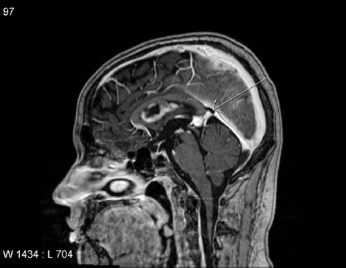
In May an increased level of AFP in serum was detected. In June MRI showed diffuse tumour recurrence in the pineal gland, in the lateral ventricles, the third ventricle, and an indication of meningeal involvement of both temporal poles (). Intrathecal methotrexate treatment combined with systemic high-dose methotrexate was given.
In August 2009 the patient's clinical condition had worsened. He suffered from intense headache and explosive vomiting. The sensibility loss on the right side of the face and the facial paralysis had progressed. The patient had trouble controlling the right-sided extremities and showed a staggering walk. All efficacious treatment options with a documented effect had been tried. The headache, nausea and vomiting was relieved with solu-medrol. The cerebral condition deteriorated and his memory and walk worsened progressively. CT-scan of the brain showed pronounced tumour progression and hydrocephalus, but surgical drain establishment was not possible. The patient died in late August 2009.
Discussion
Our patient is unusual in two aspects: 1) He had concomitant occurrence of intracranial GCT and carcinoma in situ (CIS) in one testicle; 2) Though he only had an intracranial GCT, an increased level of the tumour marker AFP was detected in the serum but not in the CSF.
Dual occurrence of pineal germinoma and unilateral testicular seminoma has previously been reported in only three cases. In two of these cases, the gonadal seminoma occurred many years after the pineal germinoma. According to Peat et al., the intracranial and gonadal germinoma represent two primary germ cell tumours. They report a case with a time interval of 16 years between diagnosis of a testicular seminoma and a cerebral germinoma. It is highly improbable that the original pineal tumour was a metastasis from a seminoma appearing 16 years later. Correspondingly, it seems unlikely that the seminoma was a distant metastasis originating from a primary intracranial germinoma, as no other extracranial metastases were evident in the patient.
Testicular biopsy in our patient revealed CIS in one testicle. This precursor of a germ cell tumour has the potential of developing into the different subtypes of testicular germ cell tumours, including seminoma. Referring to the theoretical explanations above, it seems unlikely that the CIS has originated from the intracranial GCTs when no other extracranial tumour was detected. It has been proposed that the dual occurrence of two primary GCTs in one patient either represent a mutation in the germ cell line, or a result of oncogenic factors affecting gonadal and extragonadal germ cells simultaneously [Citation2,Citation3].
As far as we are aware, the finding of tumour markers in the blood but not in the CSF in patients with icGCT has only been reported in one case previously. Ozbey et al. described a female patient diagnosed with an intrasellar germinoma who had a normal β-hCG concentration in the CSF but an increased level of β-hCG in the serum. CT-scan and MRI of chest, abdomen and pelvis were normal. The tumour marker level decreased in the peripheral blood after radiotherapy treatment of the tumour region. The authors suggested an explanation, that some intracranial germinomas are not in direct connection with the subarachnoidal space and tumour markers are therefore not secreted to the CSF. As the tumour communicates with the systemic circulation via hypophyseal venous circulation this could explain the increased level of tumour markers in the blood but not in CSF [Citation4].
The patient described in our case story had increased concentrations of AFP initially in the peripheral blood but normal concentrations in the spinal fluid. CT-scan of thorax and abdomen did not reveal extracranial tumours that could explain the elevated tumour marker levels in serum. After the first treatment course, MRI showed remission of tumour and serum levels of AFP was normalised. This supports the hypothesis, that the intracranial GCT was responsible for the elevated AFP level in serum, since the disappearance of the tumour correlated with decreased tumour marker concentrations in the peripheral blood. It is obtrusive that the AFP concentration is increased in serum and not in CSF. However, it cannot be ruled out that a small germ cell tumour in, for instance the retroperitoneum, not identified by the mentioned investigation, could be responsible for the AFP production. In the case of Ozbey et al. this is obviously not the case, as the patient only received treatment against the intracranial tumour.
The co-existence of an intracranial germ cell tumour with carcinoma-in-situ in a testicle support the theory that these conditions are part of a general syndrome, e.g. Testicular dysgenesis syndrome (TDS) as suggested by Skakkebæk et al. TDS is a result of disruption of embryonal programming and gonadal development during foetal life due to adverse environmental influences [Citation5].
Declaration of interest:
The authors report no conflicts of interest. The authors alone are responsible for the content and writing of the paper.
References
- Echevarria ME, Fangusaro J, Goldman S. Pediatric central nervous system germ cell tumours: A review. Oncologist 2008;13:690–9.
- Ho CL, Deruytter MJ. Dual occurence of pineal germinoma and testicular seminoma. Pediatr Neurosurg 2006;42: 38–44.
- Peat DS, Trowell JE. Testicular seminoma in a patient with pineal germinoma. J Clin Pathol 1994;47:771–2.
- Ozbey N, Sencer A, Tanyoloc S, Kurt R, Sencer S, Bilgic B, . An intrasellar germinoma with normal cerebrospinal fluid β-hCG concentrations misdiagnosed as hypophysitis. Hormones 2006;5:67–71.
- Skakkebæk NE, Rajpert-De Meyts E, Main KM. Testicular dysgenesis syndrome: An increasingly common developmental disorder with environmental aspects: Opinion. Hum Reprod 2001;16:972–8.

