
Figures & data
Figure 1. Flow chart of all subject episodes grouped according to the index test and reference standard. Group names are marked in bold.
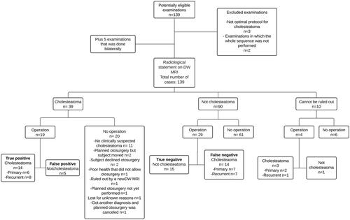
Table 1. Contingency table with otosurgery as the reference standard and non-EPI DW MRI as the index test.
Figure 2. Total number of executed non-EPI DW MRI per year separated into each radiological diagnosis. non- EPI DW MRI: Non-Echo Planar Imagining Diffusion Weighed Magnetic Resonance Imaging.
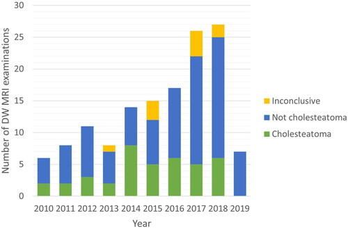
Figure 3. Time to otosurgery from the date of non-EPI DW MRI for all operated groups. non- EPI DW MRI: Non-Echo Planar Imagining Diffusion Weighed Magnetic Resonance Imaging.
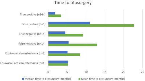
Table 2. Sex, type of disease and follow-up for all operated groups.
Table 3. Sensitivity, specificity, predictive values, and likelihood ratios for non-EPI DW MRI to detect cholesteatoma.
Data availability statement
The data that support the findings of this study are available from the corresponding author, [EK], upon reasonable request.