Figures & data
Figure 1. Stigma (a, d, g, j), style (b, e, h, k) and nucellar tissue (c, f, i, l) of female flower with DAPI (a–f) and TUNEL (g–l) assay. (a–c) Prior to PCD, chromatin is evenly distributed in the nuclei. (g–i) Prior to PCD, TUNEL is negative. (d–f) Condensed chromatin (arrow) in nuclei in advanced stage of PCD. (j–l) TUNEL is positive (arrow) in advanced stage of PCD. sg, stigma; st, style; nu, nucellus. Scale 10 µm (a–f) and 50 µm (g–l).
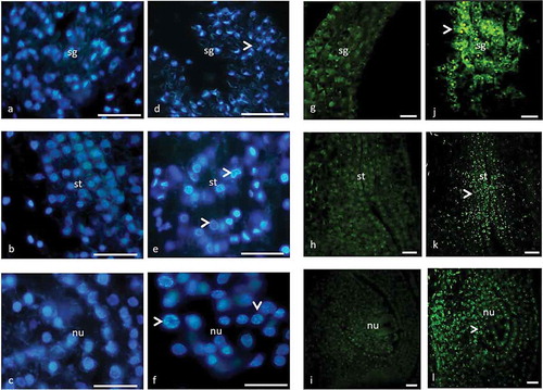
Figure 2. Stigma (a, d, g, j), style (b, e, h, k) and nucellar tissue (c, f, i, l) of male flower with DAPI (a–f) and TUNEL (g–l) assay. (a–c) Prior to PCD, chromatin is evenly distributed in the nuclei. (g–i) Prior to PCD, TUNEL is negative. (d–f) Condensed chromatin (arrow) in nuclei in advanced stage of PCD. (j–l) TUNEL is positive (arrow) in advanced stage of PCD. sg, stigma; st, style; nu, nucellus. Scale 10 µm (a–f) and 50 µm (g–l).
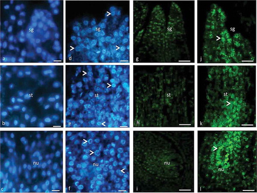
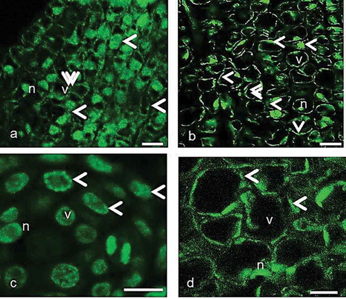
Figure 3. The organization of immunofluorescently labeled microtubules in the pistil cells of female, gall and male flowers prior to programmed cell death visualized by fluorescence microscopy (c) and laser confocal microscopy (a, b, d). The arrowheads show fluorescence signal on the cortical region, around the nuclei and vacuoles. The double arrowheads show thick fluorescent aggregates. Scale 10 μm.
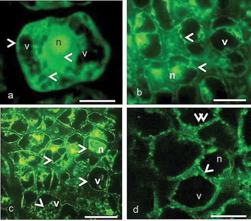
Figure 4. The organization of immunofluorescently labeled microtubules in the pistil cells of female, gall and male flowers in the course of programmed cell death visualized by laser confocal microscopy. (a–c) The arrowheads show fluorescence signal on the cortical region, around the nuclei and vacuoles. The double arrowheads show thick fluorescent aggregates. (d) The randomly organized cortical MT network. n, nucleus; v, vacuole. Scale 10 μm.