
Figures & data
Figure 1. Histology slide stained with Hematoxylin and Eosin (H&E) showing prominent perivascular cuffing (arrow) with lymphocytes, few plasma cells and some macrophages and formation of multiple glial nodules (star). Quadrigeminal bodies, mesencephalon. Scale bar: 100 µm.
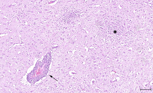
Table 1. RT-qPCR results for tick-borne encephalitis virus (TBEV).
Figure 2. Detection of tick-borne encephalitis virus (TBEV) RNA in the brain of three Dalmatian puppies by in situ hybridization (ISH). Positive hybridization signals shown as red granular staining can be seen in the claustrum of puppy ‘S19-1708’ (a). Positive signals for TBEV RNA can be seen in the thalamus of puppy ‘S19-1722’ (B) and in the thalamus of puppy ‘S19-1723’ (C). No in situ detection of TBEV RNA in the brain of a healthy young dog serving as a negative control (D). Scale bars: 100 µm.
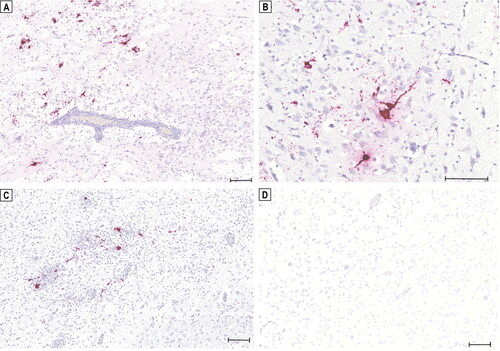
Figure 3. Phylogenetic comparison of whole genome sequences of representative tick-borne encephalitis virus (TBEV) strains from the three main subtypes. Representative full-length sequences were obtained from NCBI GenBank with corresponding accession numbers shown in brackets. Colors indicate the three different TBEV subtypes (green: European subtype [TBEV-Eu], red: Far Eastern subtype [TBEV-Fe], blue: Siberian subtype [TBEV-Sib]). The obtained consensus sequence of puppy ‘S19-1723’, TBEV-Eu-Switzerland-2019-Thurgau (bold), clusters with the European subtypes.
![Figure 3. Phylogenetic comparison of whole genome sequences of representative tick-borne encephalitis virus (TBEV) strains from the three main subtypes. Representative full-length sequences were obtained from NCBI GenBank with corresponding accession numbers shown in brackets. Colors indicate the three different TBEV subtypes (green: European subtype [TBEV-Eu], red: Far Eastern subtype [TBEV-Fe], blue: Siberian subtype [TBEV-Sib]). The obtained consensus sequence of puppy ‘S19-1723’, TBEV-Eu-Switzerland-2019-Thurgau (bold), clusters with the European subtypes.](/cms/asset/53b8aa3d-d6bf-4722-a724-46927d54d76a/tveq_a_2338385_f0003_c.jpg)
Supplemental Material
Download Zip (138.6 KB)Data availability statement
Raw sequencing reads generated and analysed are available at the National Center for Biotechnical Information (NCBI) sequence read archive under the Accession Number PRJNA1014446. The genome of TBEV-EU-Switzerland-2019-Thurgau is available at NCBI GenBank under the Accession Number OR523238.