
Figures & data
Figure 1. The approximate locality on the Waoku Coach Road Track (red circle), Parataiko Range, Northland, Aotearoa – New Zealand where the two specimens of Mecodema haakuturi sp. nov. were collected by the late John Ward, Canterbury Museum, Christchurch.
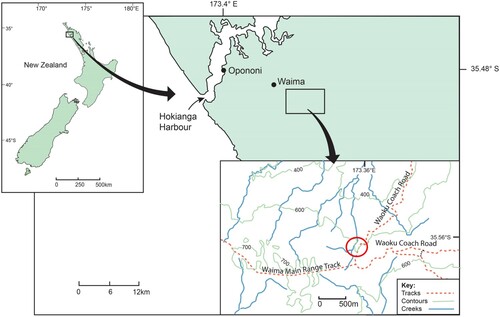
Table 1. Described regionally endemic Mecodema species (in alphabetical order), the type locality and the ranges within the Northland (ND) region as per Crosby et al. (Citation1998), New Zealand.
Figure 2. Male genitalia (aedeagus) structures (detached) of Mecodema haakuturi: (AL) aedeagal lobe, length 4.6 mm; (LP) left paramere (detached from AL), length 3.65 mm; (RP) right paramere (detached from AL), length 3.4 mm (EP) endophallus, length 3.9 mm (everted from AL). The Y-sclerite contains the sclerotised central spicule and associated structures (Photographs by author: DSS) [CMNZ specimen 2007.163.10642].
![Figure 2. Male genitalia (aedeagus) structures (detached) of Mecodema haakuturi: (AL) aedeagal lobe, length 4.6 mm; (LP) left paramere (detached from AL), length 3.65 mm; (RP) right paramere (detached from AL), length 3.4 mm (EP) endophallus, length 3.9 mm (everted from AL). The Y-sclerite contains the sclerotised central spicule and associated structures (Photographs by author: DSS) [CMNZ specimen 2007.163.10642].](/cms/asset/a34b1b61-1ea3-4421-a28a-d394d37f9716/tnzz_a_2334022_f0002_oc.jpg)
Figure 3. Mecodema haakuturi sp. nov. plate: A, dorsal habitus; B, habitus illustration; C, ventral habitus. The scale bar under the habitus is the width across the widest point of the elytra of the specimen used for this illustration. Illustration by Vivian Ward, School of Biological Sciences, University of Auckland (Photographs by author: DSS) [CMNZ specimen 2007.163.10642].
![Figure 3. Mecodema haakuturi sp. nov. plate: A, dorsal habitus; B, habitus illustration; C, ventral habitus. The scale bar under the habitus is the width across the widest point of the elytra of the specimen used for this illustration. Illustration by Vivian Ward, School of Biological Sciences, University of Auckland (Photographs by author: DSS) [CMNZ specimen 2007.163.10642].](/cms/asset/2f19be62-bbf2-4b23-a4ed-6aeb3fada705/tnzz_a_2334022_f0003_oc.jpg)
Figure 4. Ventral view of a Mecodema specimen showing specific morphological structures, excluding taxonomic structures indicated in detail figures, used in the species descriptions. VLF = ventrite lateral foveae; VSP = ventrite setose punctures; MTC = metacoxa; MTVP = metaventrite process (with carina); MSC = mesocoxa; PC = procoxa; PS = prosternum; G = gena; PES = proepisternum; MSE = mesepisternum; MTE = metepisternum; V1–V6 = ventrites 1–6 (ventrites 1–3 may be fused); M = midline (dashed line, not a taxonomic structure) [Reproduced from Seldon & Buckley (Citation2019) with permission from copyright holder, Zootaxa].
![Figure 4. Ventral view of a Mecodema specimen showing specific morphological structures, excluding taxonomic structures indicated in detail figures, used in the species descriptions. VLF = ventrite lateral foveae; VSP = ventrite setose punctures; MTC = metacoxa; MTVP = metaventrite process (with carina); MSC = mesocoxa; PC = procoxa; PS = prosternum; G = gena; PES = proepisternum; MSE = mesepisternum; MTE = metepisternum; V1–V6 = ventrites 1–6 (ventrites 1–3 may be fused); M = midline (dashed line, not a taxonomic structure) [Reproduced from Seldon & Buckley (Citation2019) with permission from copyright holder, Zootaxa].](/cms/asset/2c0076a2-15fa-4845-93ca-ec222a1d3872/tnzz_a_2334022_f0004_ob.jpg)