
Figures & data
Figure 1. Structure of salt-secreting modified trichomes in recretohalophytes. A. A salt bladder complex includes an epidermal bladder cell (EBC) and a stalk cell in C. album (from Zhang et al. (Citation2022) and only EBC in M. crystallinum (from Oh et al. (Citation2015); scalebar: 0.5 mm); B. Salt gland structures include single salt gland in O. coarctata (Koteyeva et al., Citation2022; n, nucleus; scalebar: 10 µm), bicellular salt gland in Chloris gayana Kunth (Oi et al., Citation2012; BC, basal cell; BR, base region of basal cell; Ca, cavity; CC, cap cell; Cu, cuticular layer; E, epidermal cell; M, mesophyll cell, n, nucleus; NR, neck region of basal cell; Va, valleculae; arrows: plasmodesmata) and multicellular salt gland in L. bicolor (Yuan et al., Citation2015; SC, secretory cell; AC, accessory cell; IC, inner cup cell; OC, outer cup cell; CC collection cell; scalebar: 5 µm). Modified from: A - Zhang et al. (Citation2022) and Oh et al. (Citation2015); B – Koteyeva et al. (Citation2022), Oi et al., (Citation2012) and Yuan et al. (Citation2015) with permission from John Wiley and Sons; Springer Nature; and University of Chicago Press.
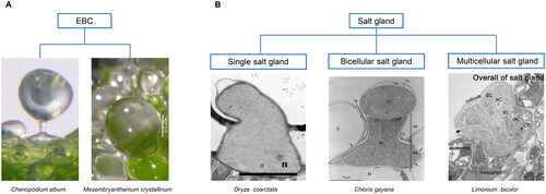
Table 1. Genes involved in formation of salt bladders or glands in recretohalophytes.
Figure 2. Maximum likelihood tree of GL2 (A), TTG2 (B) and GL3 (C) proteins from glycophytes and recretohalophytes. Genes in glycophytes and recretohalophytes were marked by solid circle. The percentage of trees in which the associated taxa clustered together is shown next to the branches. The tree is drawn to scale, with branch lengths measured in the number of substitutions per site. Evolutionary analyses were conducted in MEGA X.
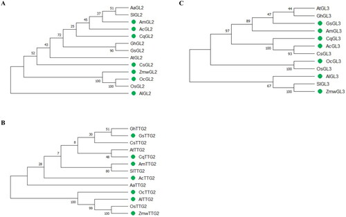
Table 2. Homologs of trichome development-related genes in glycophytes and recretohalophytes.
Figure 3. Confocal imaging of Na+ fluorescence using CoroNa Green AM in salt glands of A. lagopoides under control and 300 mM NaCl for 3 days.
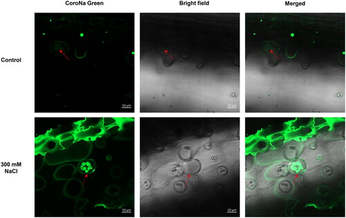
Table 3. Genomes-sequenced of recretohalophytes with salt bladders or salt glands until 2023.