Figures & data
Figure 1. Chemical structure of (A) tamoxifen citrate and (B) silk fibroin.
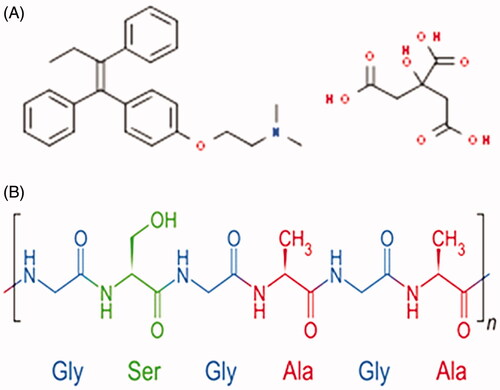
Table 1. Physical characterization of blank SF-NPs, TC-SF-NPs, and lyophilized TC-SF-NPs.
Figure 2. Surface morphology of TC-SF-NPs. (A) SEM image of TC-SF-NPs at X 100,000 (B) TEM of TC-SF-NPs. TC-SF-NPs: tamoxifen citrate silk fibroin nanoparticles; SEM: scanning electron microscopy; TEM: transmission electron microscopy.
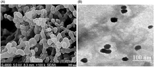
Figure 3. Physicochemical characterization of TC-SF-NPs. (A) Fourier transform infrared spectra. (B) X-ray diffraction spectra. TC-SF-NPs: tamoxifen citrate silk fibroin nanoparticles.
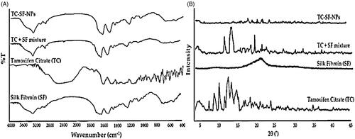
Figure 4. In vitro release study of free TC and TC-loaded SF-NPs at different pH. TC: tamoxifen citrate; TC-SF-NPs: tamoxifen citrate silk fibroin nanoparticles.
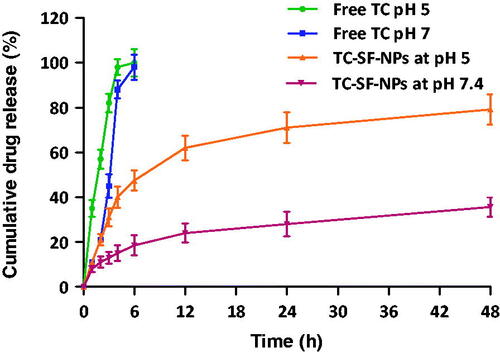
Table 2. Stability study of optimized TC-SF-NPs at different temperatures and the humidity condition.
Figure 5. In vitro cytotoxicity of TC-SF-NPs against breast cancer cell lines. (A) MCF-7 and (B) MDAMB-231 cells were incubated with different concentrations (25–400 μg/ml) of free TC, SF-NPs or TC-loaded SF-NPs for 72 h. Untreated cells served as controls. The dose of TC in TC-SF-NPs group was equivalent to that of free TC group. Cell viability was assessed by MTT assay. Data represent mean ± SD. #p < .05 vs. TC-SF-NPs, ***p < .005 vs. SF-NPs. TC: tamoxifen citrate; SF-NPs: silk fibroin nanoparticles; TC-SF-NPs: tamoxifen citrate silk fibroin nanoparticles.
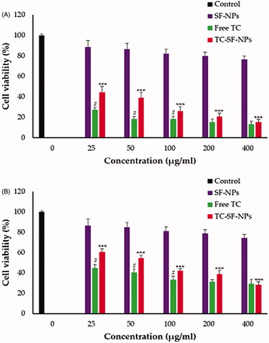
Figure 6. Cellular uptake of TC-SF-NPs by MCF-7 and MDAMB-231 cell lines. (A) Confocal microscopy images of MCF-7 and MDA-MB-231 cells treated with PI-labeled TC-SF-NPs for 24 and 48 h. (B) Flow cytometry quantitative analysis showing percentage of uptake of PI-labeled TC-SF-NPs by MCF-7 and MDA-MB-231 cells following 24 h incubation. Data are expressed in mean ± SD. **p < .01. TC: tamoxifen citrate; TC-SF-NPs: tamoxifen citrate silk fibroin nanoparticles.
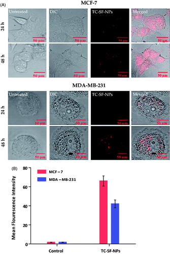
Table 3. Effect of free TC and TC-loaded SF-NPs on the redistribution of growth-arrested MCF-7 and MDA-MB-231 cells in the different phases of the cell cycle.
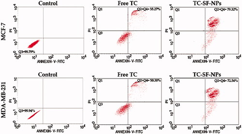
Figure 7. Cellular apoptosis of breast cancer cells treated with TC-loaded SF-NPs. MCF-7 and MDA-MB-231 cells (3 × 10⁵ cells/well) were treated with either free TC or TC-SF-NPS for 24 h. Cellular apoptosis was evaluated by flow cytometry using Annexin V-FITC staining. TC: tamoxifen citrate; TC-SF-NPs: tamoxifen citrate silk fibroin nanoparticles.