
Figures & data
Figure 1. Overview of the reliability study design.
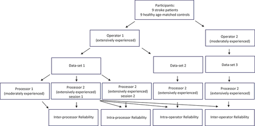
Table 1. Overview of in- and exclusion data for stroke patients and healthy age-matched controls.
Figure 2. Overview of acquisition and processing of 3DfUS.
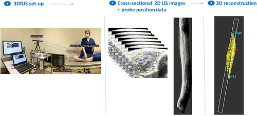
Figure 3. Participant flow diagram.
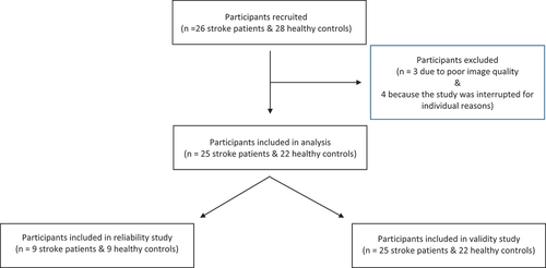
Table 2. Demographic and anthropometric information of participants. Medians (inter quartile ranges) or frequencies are given.
Table 3. Clinical scales for stroke patients & healthy subjects.
Table 4. Overview of reliability results for tibial anterior normalized muscle volume, normalized muscle length and echo-intensity for stroke patients and healthy controls.
Table 5. Comparisons between leg of stroke patients (affected/non-affected, with/without SMO, with/without TA paresis, with/without contracture) versus left leg of healthy controls (mann-whitney U test).
Table 6. Comparisons between affected versus non-affected leg in stroke patients & between left versus right leg in healthy subjects (Wilcoxon signed-rank test).