
Figures & data
Figure 1. Flowchart of the study cohort.
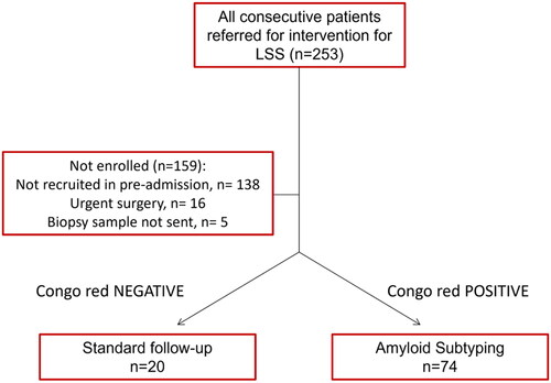
Table 1. Characteristics of the study cohort, % (N).
Figure 2. Histology of resection specimens showed fragments of soft tissue and ligamentum flavum (A). Variable amounts of amyloid were found in the ligamentum flavum (B). At higher magnification, a homogenous eosinophilic material was noted after haematoxylin and eosin staining (asterisk; C), which showed a typical yellow-green birefringence in polarized light after Congo red staining (D). These deposits immunoreacted with antibodies directed against transthyretin (E) and complement 9 (F). Haematoxylin and eosin staining (A, C); Congo red staining viewed in fluorescence microscopy (B); Congo red staining viewed in polarization microscopy (D), anti-transthyretin-immunostaining (E); anti-complement 9-immunostaining (F). Barr denotes 1 mm (A) and 50 µm (C, E, F). Original magnifications 0.56-fold (A, B); 200-fold (C–F). (G, H) Exemplary images of ligamentum flavum measurement on axial T2-weighted MR images (L2–L3 level), in a patient without (G) and with (H) proven amyloid deposition at histopathology. The corresponding measurements are given for each side in mm.
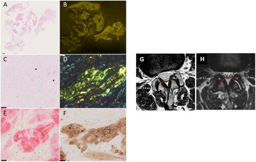
Figure 3. Box plots showing association between age, mean ligamentum flavum thickness for each level and amyloid detection. *p < .05; **p < .01.
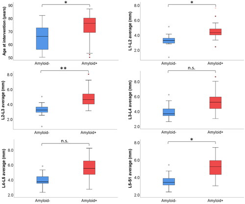
Figure 4. Forest plot showing odds ratio and 95% confidence intervals (CIs) of continuous variables (A) and binary variables (B) for the univariate logistic regression. Bar plot on the right indicates p values after multiple comparison. The red line indicates significance (FDR <0.05). Intervention for previous CTS was excluded for absence of amyloidosis in either group.
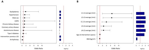
Table 2. Histopathology reports of the amyloid + cohort.
Table 3. Association between age, mean ligamentum flavum thickness and amyloid detection.
Table 4. Association between comorbidities and amyloid detection.
Supplemental Material
Download MS Power Point (176.2 KB)Supplemental Material
Download MS Word (13.9 KB)Data availability statement
Single patient data available upon motivated request by email to the corresponding author.