
Figures & data
Figure 1. (a and b). Best ROC values of fetal distress predicted by the fetal middle cerebral artery resistance index (MCA-RI) and cerebroplacental ratio 3 (CPR-3); (c and d). Best ROC values of small for gestational age (SGA) predicted by MCA-RI and CPR-3. X axis refers to 100%-Specificity%; Y axis refers to Sensitivity%.
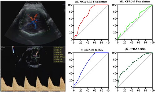
Figure 2. Mean and standard deviation were used to describe the fetal middle cerebral artery (MCA) and umbilical artery (UA) blood flow in the three different periods (20–24 weeks, 29–32 weeks and 35-40 weeks. (A). MCA-PI and UA-PI; (B). MCA-RI and UA-RI; (C). MCA-S/D and UA-S/D; (D). MCA-Vm and UA-Vm.
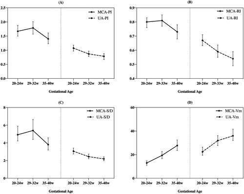
Table 1. The ultrasound detection of the fetal middle cerebral artery, umbilical artery blood flow of the normotensive controls group, GH group and PE/EC group.
Table 2. ROC values of fetal distress and small for gestational age predicted by the ultrasound detection of the fetal middle cerebral artery blood flow and CPR.