
Figures & data
Figure 1. Results of imaging studies performed at 31 weeks. (A,B) Ultrasound appearance of the pregnancy, and the molar component. (C) Sagittal MRI image. (D) Transverse MRI image.
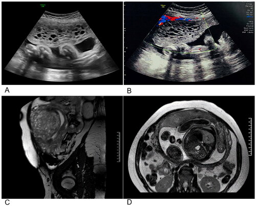
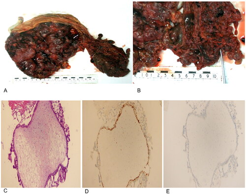
Figure 2. Pathology of the patient’s placenta. (A,B) Gross pathological assessment following delivery. A placenta from a normal pregnancy is presented adjacent to the complete molar tissues. (C) Hematoxylin-eosin staining results of the complete hydatidiform mole. (D) Ki-67 expression in the complete hydatidiform mole (+). (E) p57 expression in the complete hydatidiform mole (-).
Data availability Statement
All data analyzed during this study are included in the published article.