
Figures & data
Figure 1. Flow chart of the study. VSD: ventricular septal defect.
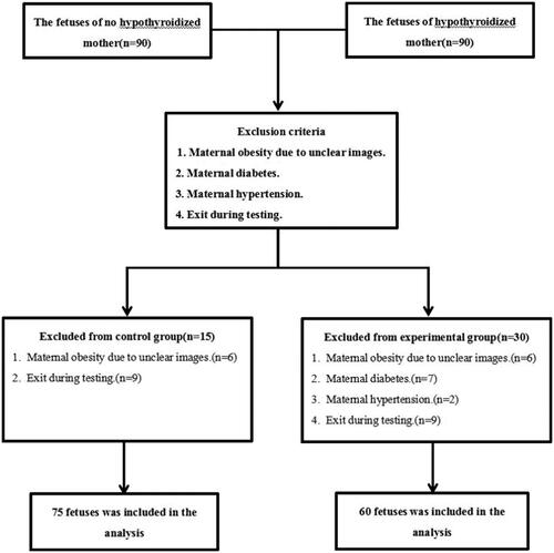
Figure 2. The diastolic and systolic diameters of left and right ventricleswere measured by M-mode echocardiography and FS were calculated. A is a 25-weeks normalfetus and B is a 25-weeks fetus in a pregnant woman with hypothyroidism. The RV FS of Bfetus was lower than that of A fetus.
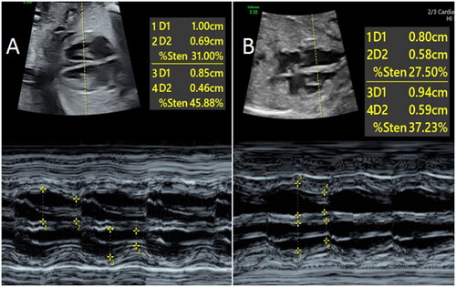
Figure 3. Schematic diagram of calculating Tei index. The time (a) between the end of mitral A wave to the beginning of E wave in the next cardiac cycle, and the duration (b) of aortic ejection were measured after the Doppler spectra of LV inflow and outflowtract were obtained. Tei index = (a–b)/b. LVOT: Left ventricular outflow tract.
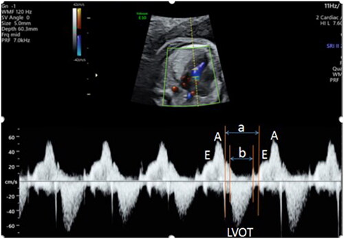
Table 1. Clinical data for hypothyroidism and control groups.
Table 2. Comparison of fetal EIF.
Table 3. Comparison of fetal myocardial thickness between hypothyroidism and control group.
Table 4. Comparison of fetal heart systolic function, ductus venosus, and pulmonary vein Doppler variables.
Table 5. Comparison of left and right ventricular Tei index.