
Figures & data
Figure 1. Glycolysis. On the left, glycolytic enzymes (blue boxes) and their products are shown representing the 10 steps of glycolysis. Steps using or generating ATP are denoted in green. On the right, the contributions of glycolysis to biosynthetic pathways. Pyruvate feeds into the TCA cycle and supports mitochondrial oxidative phosphorylation (bottom right). Examples of the distribution of select endogenous glycolysis enzymes detected through immunocytochemistry along the axons of embryonic chicken sensory neurons are shown. Enzymes are labeled in green and actin filaments in red. The examples shown are from our laboratory. Figure prepared using Biorender.com.
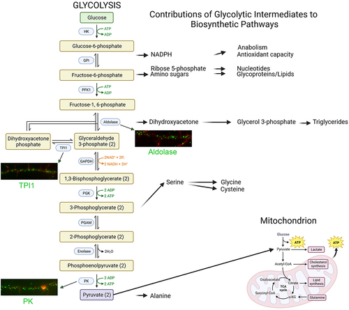
Figure 2. Glycolysis as a localized bioenergetic source along axons. (a) “On-board” glycolysis. Glycolytic enzymes are associated with fast transport vesicles along axons. The vesicle associated enzymes then generate ATP that is used by the vesicle associated motor proteins to move vesicles along microtubules. In contrast, mitochondria power their own transport by oxidative phosphorylation. (b) During cannabinoid induced axon retraction glycolytic enzymes are recruited to myosin II, the main ATPase mechanoenzyme that powers cellular contractility through actin filaments. The Myosin II associated glycolytic enzymes then generate ATP that is used by myosin II to power actomyosin contractility and drive axon retraction. Figure prepared using Biorender.com.
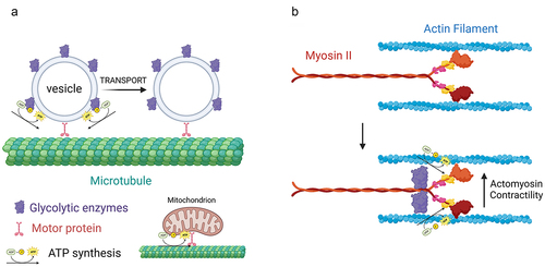
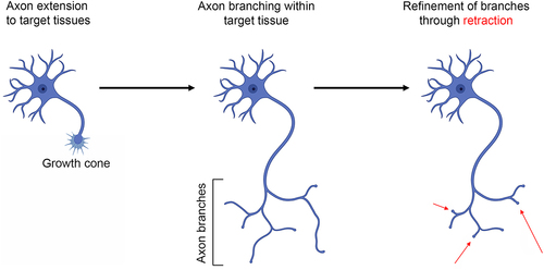
Figure 3. Overview of axon development. Axons initially form from the cell bodies, develop a growth cone, and extend to their target tissue. Within target tissues, or along the way, the axons generate collateral branches. Finally, during the period of refinement, supernumerary axon branches are pruned/retracted as a component of the ongoing establishment of synaptic circuitry. Figure prepared using Biorender.com.
Data availability statement
This is a review article and contains no data.