Figures & data
Table 1. Details concerning the SERS substrates and their preparation.
Figure 1. (a) SEM image of the annealed 1 nm nominal thickness Au-coated silicon wafer (26 nm average diameter). Dotted circle indicates approximately the size of the laser spot for Raman/SERS excitation. (b) Particle size distribution for the sample shown in (a). Average was performed on ~2000 particles. (c and d) SEM images of the <1 nm (5 nm) and 2 nm (104 nm) nominal thickness Au-coated silicon wafers respectively. Scaling bar appearing in (a) applies also on (c) and (d).
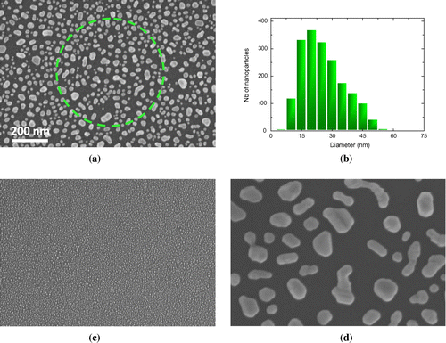
Figure 2. Reflectance spectra in the visible spectral region from the silicon wafers with average particle diameter of 5, 26 and 104 nm. Arrows designate the wavelengths of the two lasers used for the collection of the SERS spectra.
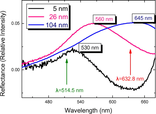
Figure 3. SERS spectra recorded from the dewetted substrates with average Au-island diameter 26 nm. The spectra correspond to the five different concentrations of the solutions deposited on them. The SERS spectrum from a colloidal SERS substrate is plotted on top.
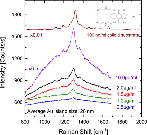
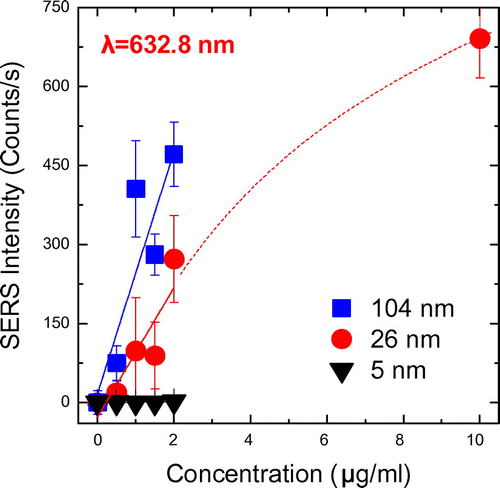
Figure 4. SERS intensity as a function of MTX concentration in the solution deposited on the dewetted Au-coated silicon wafers. Linear fits are shown up to 2 μg ml−1 while an estimated dotted curve is drawn towards the 10 μg ml−1 for the substrates with 26 nm sized Au-islands.