
Figures & data
Figure 1. Overview of Mtb infection. Mtb enters the human body through the airway where it engages the innate immune system within the alveolar space. Macrophages and dendritic cells ingest the bacteria, recruiting new cells and activating adaptive immunity. Together, the innate and adaptive immune systems collaborate to eradicate the bacteria or restrict its active replication within a granuloma. Active tuberculosis occurs either after primary infection or after reactivation due to immunodeficiency, leading to symptomatic disease and transmission to a new host to start a new infection cycle.
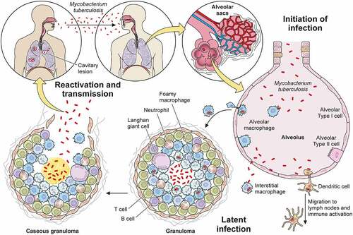
Table 1. Non-protein virulence factors of Mtb.
Figure 2. Non-protein virulence factors of Mtb. Schematic of Mtb lipids, glycolipids, glycans, and nucleic acids that contribute to virulence. TDM: trehalose dimycolate, SL-1: sulfolipid-1, PGL: phenolic glycolipid, MA: mycolic acid, PAT: pentaacyl trehalose, PDIM: phthiocerol dimycocerosates, ManLAM: mannosylated lipoarabinomannan.
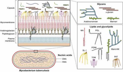
Figure 3. Protein virulence factors of Mtb. Schematic representation of Mtb interactions within infected macrophages highlighting (i) toll-like receptors, (ii) phagosome arrest and escape, (iii) autophagy subversion, (iv) cell death modulation, and (v) cytokine production. Each box contains representative Mtb virulence factors that engage host cell biologic processes detailed in the text.
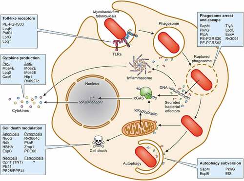
Table 2. Protein virulence factors of Mtb.