Figures & data
Figure 1. Schematic diagram illustrating outer inner membrane vesicles (OIMV) of Gram negative bacteria (containing cytoplasmic proteins in addition to the periplasmic space proteins in outer membrane vesicles), cytoplasmic membrane vesicles (CMV) of Gram positive bacteria, fungal extracellular vesicles and exosomes. SOD – superoxide dismutase, PG – peptidoglycan, SabA – sialic acid-binding adhesin, VacA – vacuolating cytotoxin, lnlB – Listeria monocytogenes protein, BlaZ – beta lactamase, Hsp – heat shock protein.
Table 1. List of major extracellular vesicle cargo reported from bacteria and fungi.
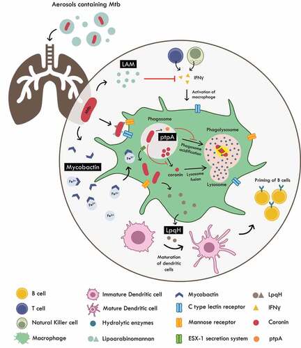
Figure 2. Host pathogen interactions of M. tuberculosis. Mtb is internalized by the macrophages via host receptors such as C – type lectin and mannose receptors that recognize LAM and LpqH respectively. Once internalized, the pathogen is subjected to intra-phagosomal processing. Mtb survives by producing molecules such as ptpA (that prevents phagosome acidification) and inhibition of secretion of coronin (important for lysosome – phagosome fusion). Mtb also induces T cell anergy and hampers the production of IFNγ by T cells and natural killer cells rendering them unable to activate the macrophage. LpqH released from lysis of Mtb are utilized by host to induce maturation of dendritic cells which then travel to lymph nodes to prime B cells for clearing of Mtb. Mtb is able to survive iron starvation by enhancing secretion of vesicles charged with mycobactin (siderophore). These key molecules can potentially serve as efficacious diagnostic biomarkers of TB.