
Figures & data

Figure 1. Stability of different types of liposomes. Data are represented as mean ± SD (n = 3).
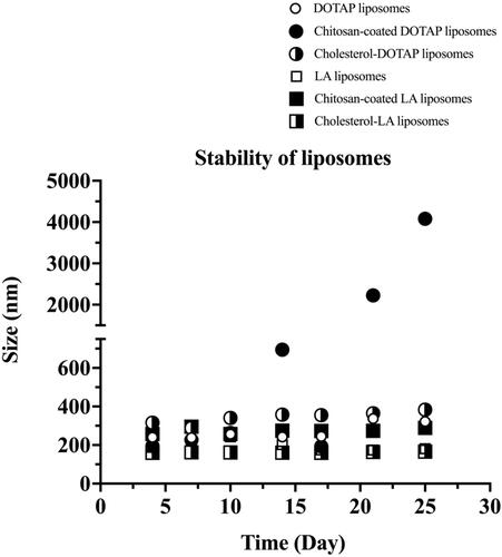
Table 1. Characterization of the size and zeta potential for different types of liposomes.
Figure 2. Transmission electron microscopy of (a) DOTAP liposomes, (b) chitosan–coated DOTAP liposomes, (c) cholesterol–DOTAP liposomes, (d) LA liposomes, (e) chitosan–coated LA liposomes and (f) cholesterol–LA liposomes with a magnification of ×25,000. The chitosan coating layer is indicated by arrows.
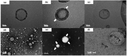
Figure 3. The secretion of isolated EVs from immortalized adipose-derived mesenchymal stem cells after being treated with different dosages and types of liposomes: (a) DOTAP liposomes, (b) chitosan–coated DOTAP liposomes, (c) cholesterol–DOTAP liposomes, (d) LA liposomes, (e) chitosan–coated LA liposomes and (f) cholesterol–LA liposomes. Data are represented as mean ± SD (n = 3), **p < .01, ***p < .001.
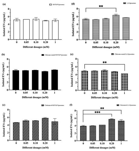
Figure 4. The percentage of cell viability after being treated with different dosages and types of liposomes. Data are represented as mean ± SD (n = 3), *p < .05, **p < .01, ***p < .001.
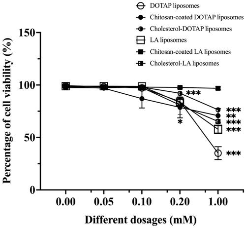
Figure 5. Fluorescence microscopy of (a) LA liposomes before and after being separated from the conditioned medium and (b) cholesterol–LA liposomes before and after being separated from the conditioned medium. The green spherical shape represents the presence of liposomes in conditioned medium before separation.
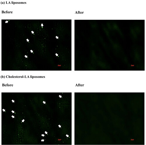
Table 2. Characterization of the size and zeta potential for different types of liposomal-EVs with wound healing properties.
Figure 6. Transmission electron microscopy of (a) non-induced EVs, (b) LA liposomal-EVs and (c) cholesterol–LA liposomal-EVs. All EVs with a magnification of ×10,000 are indicated by arrows, respectively.
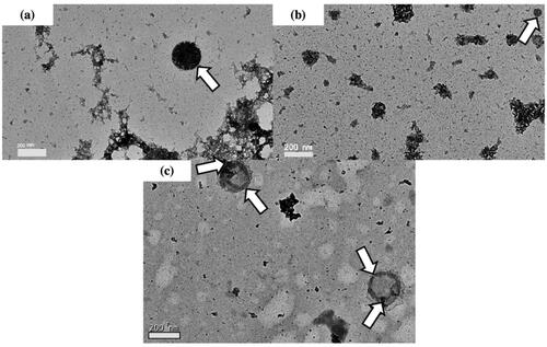
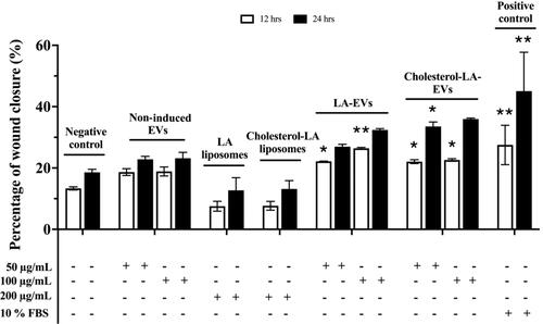
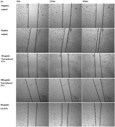
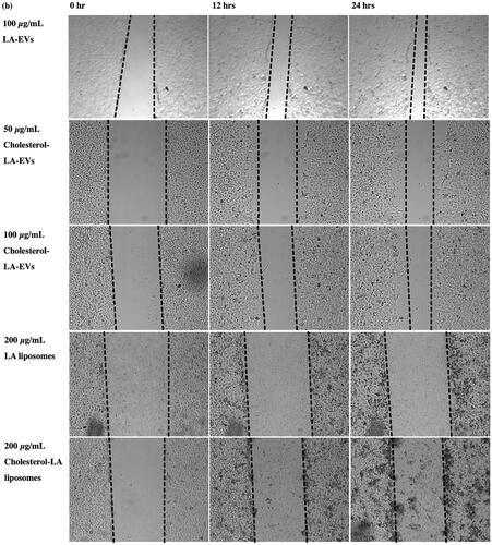
Figure 7. The percentage of wound closure in HaCaT after treatment with different dosages and types of liposomal-induced EVs. Data are represented as mean ± SEM (n = 3), *p < .05, **p < .01 .
Data availability statement
The data that support the findings of this study are available from the corresponding author upon reasonable request.