3,627
Views
5
CrossRef citations to date
0
Altmetric
Coronaviruses

The role of SARS-CoV-2 nucleocapsid protein in antiviral immunity and vaccine development

, , , &
Article: e2164219 | Received 09 Nov 2022, Accepted 28 Dec 2022, Published online: 01 Mar 2023

Figures & data

Figure 1. The structure of SARS-CoV-2 N-protein. (A). Model of SARS-CoV-2 N-protein dimer. (B). Topology diagram for SARS-CoV-2 N-protein N-terminal RNA-binding domain. (C). Superimposed structures in ribbon representation of N-protein N-terminal RNA-binding domain from SARS-CoV-2 (green), SARS-CoV (cyan), HCoV-OC43 (pink), MERS-CoV (orange), and HCoV-NL63 (blue) [Citation6]. The loops that show significant differences are highlighted by dotted circles. The AMP ligand in the HCoV-OC43 N-protein N-terminal RNA-binding domain-AMP complex is shown as a stick structure. D. Topology diagram for SARS-CoV-2 N-protein C-terminal dimerization domain. E. Superimposed structures in ribbon representation of N-protein C-terminal dimerization domain from SARS-CoV-2 (pink), SARS-CoV (cyan), MERS-CoV (orange) and HCoV-NL63 (blue). Figure adapted from [Citation6].

Figure 1. The structure of SARS-CoV-2 N-protein. (A). Model of SARS-CoV-2 N-protein dimer. (B). Topology diagram for SARS-CoV-2 N-protein N-terminal RNA-binding domain. (C). Superimposed structures in ribbon representation of N-protein N-terminal RNA-binding domain from SARS-CoV-2 (green), SARS-CoV (cyan), HCoV-OC43 (pink), MERS-CoV (orange), and HCoV-NL63 (blue) [Citation6]. The loops that show significant differences are highlighted by dotted circles. The AMP ligand in the HCoV-OC43 N-protein N-terminal RNA-binding domain-AMP complex is shown as a stick structure. D. Topology diagram for SARS-CoV-2 N-protein C-terminal dimerization domain. E. Superimposed structures in ribbon representation of N-protein C-terminal dimerization domain from SARS-CoV-2 (pink), SARS-CoV (cyan), MERS-CoV (orange) and HCoV-NL63 (blue). Figure adapted from [Citation6].

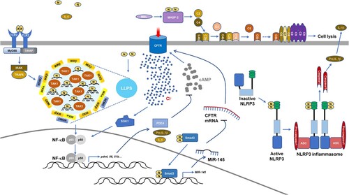
Figure 2. Induced inflammation by SARS-CoV-2 N-protein.

Figure 2. Induced inflammation by SARS-CoV-2 N-protein.

Table 1. Effects of different human-infecting coronavirus N-proteins on host cell signaling.

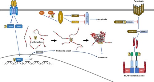
Figure 3. Modulation of cell death by SARS-CoV-2 N-protein.

Figure 3. Modulation of cell death by SARS-CoV-2 N-protein.

Table 2. SARS-CoV-2-N-based vaccines under development.