2,025
Views
1
CrossRef citations to date
0
Altmetric
Research Article

Inhalable vaccine of bacterial culture supernatant extract mediates protection against fatal pulmonary anthrax

, , , , , , , , , , , , & show all
Article: 2191741 | Received 05 Dec 2022, Accepted 08 Mar 2023, Published online: 31 Mar 2023

Figures & data

Figure 1. Characterization of B. anthracis mutants deficient in extracellular protease activity. (A) Schematic diagram of CRISPR-Cas9 genome editing in B. anthracis. (B) PCR identification of B. anthracis mutant with reduced proteolytic activity. (C) Mutant strains identified by SDS-PAGE and western blot analysis with mouse anti-PA polyclonal antibodies. (D) The relative expression levels of PA in the mutant strains analyzed by ImageJ. (E) A16R-5 and A16R-5.1 strains identified by SDS-PAGE and western blot analysis with mouse anti-PA and anti-LF polyclonal antibodies. (F) The relative expression levels of PA in A16R-5 and A16R-5.1 strains analyzed by ImageJ. (G) Survival in mice (n = 10) immunized with A16R-5 and A16R-5.1 via i.t. inoculation. (H) Volcano plots of differentially expressed proteins (DEPs) in A16R culture supernatant and CSE vaccine.
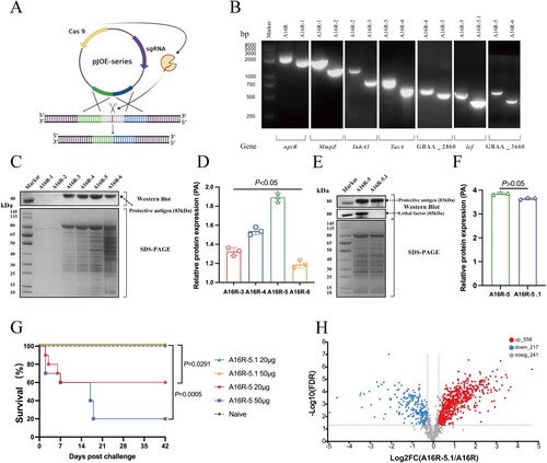
Figure 1. Characterization of B. anthracis mutants deficient in extracellular protease activity. (A) Schematic diagram of CRISPR-Cas9 genome editing in B. anthracis. (B) PCR identification of B. anthracis mutant with reduced proteolytic activity. (C) Mutant strains identified by SDS-PAGE and western blot analysis with mouse anti-PA polyclonal antibodies. (D) The relative expression levels of PA in the mutant strains analyzed by ImageJ. (E) A16R-5 and A16R-5.1 strains identified by SDS-PAGE and western blot analysis with mouse anti-PA and anti-LF polyclonal antibodies. (F) The relative expression levels of PA in A16R-5 and A16R-5.1 strains analyzed by ImageJ. (G) Survival in mice (n = 10) immunized with A16R-5 and A16R-5.1 via i.t. inoculation. (H) Volcano plots of differentially expressed proteins (DEPs) in A16R culture supernatant and CSE vaccine.

Table 1. Summary of immunization groups used in the experiment.

Figure 2. Characterization of CSE vaccine dry powder. (A) CSE dry powder and liquid analyzed by SDS-PAGE and western blot analysis with mouse anti-CSE polyclonal antibodies. (B) Immunogenicity of CSE dry powder and CSE liquid analyzed by ELISA with mice immunized with three CSE vaccines. (C) Scanning electron microscopy (SEM) images of CSE dry powder. (D) Moisture content of CSE and CpG dry powders (control). (E) VMD and (F) MMAD of aerosolized CSE dry powder.
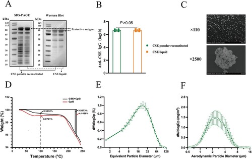
Figure 2. Characterization of CSE vaccine dry powder. (A) CSE dry powder and liquid analyzed by SDS-PAGE and western blot analysis with mouse anti-CSE polyclonal antibodies. (B) Immunogenicity of CSE dry powder and CSE liquid analyzed by ELISA with mice immunized with three CSE vaccines. (C) Scanning electron microscopy (SEM) images of CSE dry powder. (D) Moisture content of CSE and CpG dry powders (control). (E) VMD and (F) MMAD of aerosolized CSE dry powder.

Figure 3. Immune responses and LeTx neutralizing antibodies after mice were immunized with different CSE vaccines. (A) Schematic timeline of immunization, challenge, and sera and BALF collection. (B, C, D, E) Reciprocal titers of (B) IgG, (C) IgG1, (D) IgG2a, and (E) IgG2a/IgG1 antibodies to CSE in serum. (F, G) Reciprocal titers of (F) IgG and (G) IgA antibodies to CSE in BALF. (H, I) Neutralizing antibody titers to CSE in (H) serum and (I) BALF. TNA titers were determined as inflection points on antibody dilution curves and reported as effective dilution at 50% inhibition of anthrax LeTx toxicity. Values were then log10-transformed.
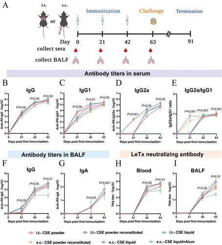
Figure 3. Immune responses and LeTx neutralizing antibodies after mice were immunized with different CSE vaccines. (A) Schematic timeline of immunization, challenge, and sera and BALF collection. (B, C, D, E) Reciprocal titers of (B) IgG, (C) IgG1, (D) IgG2a, and (E) IgG2a/IgG1 antibodies to CSE in serum. (F, G) Reciprocal titers of (F) IgG and (G) IgA antibodies to CSE in BALF. (H, I) Neutralizing antibody titers to CSE in (H) serum and (I) BALF. TNA titers were determined as inflection points on antibody dilution curves and reported as effective dilution at 50% inhibition of anthrax LeTx toxicity. Values were then log10-transformed.

Figure 4. Antigen-specific T cell immune and lymphocyte proliferation responses in mice immunized with different CSE vaccines. (A) Schematic timeline of immunization, challenge, and spleen collection. (B) T lymphocyte proliferation assay. (C, E) ELISPOT and antigen-specific Biosys Bioreader 7000 determinations of (C) IFN-γ and (E) IL-4. (D, F) Antigen-specific measurements of (D) IFN-γ and (F) IL-4.
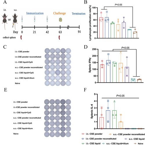
Figure 4. Antigen-specific T cell immune and lymphocyte proliferation responses in mice immunized with different CSE vaccines. (A) Schematic timeline of immunization, challenge, and spleen collection. (B) T lymphocyte proliferation assay. (C, E) ELISPOT and antigen-specific Biosys Bioreader 7000 determinations of (C) IFN-γ and (E) IL-4. (D, F) Antigen-specific measurements of (D) IFN-γ and (F) IL-4.

Figure 5. Protective efficacies of CSE vaccines against aerosolized B. anthracis spore challenges. (A) Schematic timeline of aerosol B. anthracis spore challenge and organs collection. (B, C) Survival of mice (n = 10) following (B) 100 × LD50, (C) 200 × LD50 aerosolized B. anthracis spore challenge, respectively. (D – G) Bacterial and spore loads of mice euthanized at days 2, 14, and 28 post 100 × LD50 aerosolized B. anthracis spore challenge. (D) lungs, (E) spleens, (F) livers, and (G) blood. Limits of detection (LoD) were 400 CFU for lungs, spleens, and livers and 100 CFU for blood. Challenges with 100 × LD50 B. anthracis spores were performed twice on different lots of immunized mice.
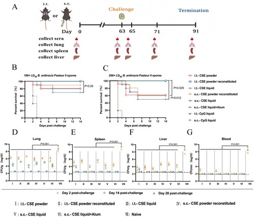
Figure 5. Protective efficacies of CSE vaccines against aerosolized B. anthracis spore challenges. (A) Schematic timeline of aerosol B. anthracis spore challenge and organs collection. (B, C) Survival of mice (n = 10) following (B) 100 × LD50, (C) 200 × LD50 aerosolized B. anthracis spore challenge, respectively. (D – G) Bacterial and spore loads of mice euthanized at days 2, 14, and 28 post 100 × LD50 aerosolized B. anthracis spore challenge. (D) lungs, (E) spleens, (F) livers, and (G) blood. Limits of detection (LoD) were 400 CFU for lungs, spleens, and livers and 100 CFU for blood. Challenges with 100 × LD50 B. anthracis spores were performed twice on different lots of immunized mice.

Figure 6. Pathological changes in tissues of CSE vaccine-immunized mice after aerosolized B. anthracis spore challenge. Mice (three per group) were euthanized day 2 post 100 × LD50 i.t. B. anthracis spore challenge. Portions of lungs, spleens, and livers were collected. (A) H&E staining of tissues from vaccinated mice. Black arrows indicate neutrophil or lymphocyte infiltration. (B) H&E staining of tissues of control mice. Red square indicates enlargement. (C) Tissue sections were evaluated as follows: 0, no pathological lesions; 1, minimal; 2, mild; 3, moderate; 4, severe.
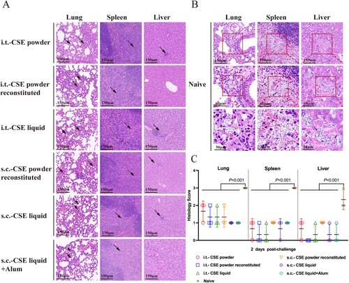
Figure 6. Pathological changes in tissues of CSE vaccine-immunized mice after aerosolized B. anthracis spore challenge. Mice (three per group) were euthanized day 2 post 100 × LD50 i.t. B. anthracis spore challenge. Portions of lungs, spleens, and livers were collected. (A) H&E staining of tissues from vaccinated mice. Black arrows indicate neutrophil or lymphocyte infiltration. (B) H&E staining of tissues of control mice. Red square indicates enlargement. (C) Tissue sections were evaluated as follows: 0, no pathological lesions; 1, minimal; 2, mild; 3, moderate; 4, severe.

Figure 7. Immune responses and relative protective efficacies of CSE vaccine and rPA. (A) Schematic timeline of immunization and challenge. (B) Reciprocal IgG titers in serum. (C, D) Reciprocal (C) IgG and (D) IgA titers in BALF. (E) Survival of mice (n = 10) against 100 × LD50 aerosolized B. anthracis spore challenge.
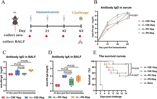
Figure 7. Immune responses and relative protective efficacies of CSE vaccine and rPA. (A) Schematic timeline of immunization and challenge. (B) Reciprocal IgG titers in serum. (C, D) Reciprocal (C) IgG and (D) IgA titers in BALF. (E) Survival of mice (n = 10) against 100 × LD50 aerosolized B. anthracis spore challenge.
Supplemental material

Supplemental Material

Download MS Word (187.9 KB)