Figures & data
Table 1. Showing the main differences between male schistosoma mansoni and S. haematobium as revealed by silver impregnation.
Figure 1. Light micrographs for AgNO3-treated male S. mansoni. (a). Nerve sensilla (NS) of dorsal surface of the middle region. (b). Venteral sucker showing ventral nerve of the sucker (VN) and venteral sucker rings (VR). (c). Anterior region of the male. Showing anteroventral connectives (AVC), cereberal ganglia (CG), oral sucker ring (OR), ring commissures (RC), venteral nerve cord (VNC) and venteral sucker (VS). (d). Dorsal surface showing nerve cell (NC).
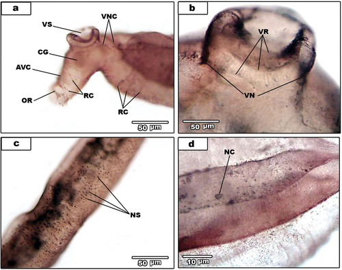
Figure 2. Light micrographs for AgNO3-treated male S. haematobium.
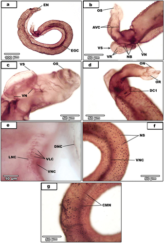
Figure 3. (A-e).Light micrographs for AgNO3-treated male S. haematobium. (a). magnification of nerve sensilla (NS). Note circular muscles nerves (black arrows). (b). dorsal surface showing tubercles (T) and ventral nerve cord (VNC). (C). male with dorsal connectives (DC) and the gynaecophoral canal (GC). (d). magnification of the posterior regionshowing dorsal connectives (DC), dorsal nerve cord (DNC) and inner dorsal nerve cord (IDNC). (e). excretory pore (EP). (f-h) light micrographs for AgNO3-treated female S. haematobium. (f). Whole female. (g). Magnification of posterior region showing ring commissures (RC) and ventral nerve cord (VNC). (3 H). magnification of anterior region (A), egg (E), ootype (OӦ) and uterus wall (UW) and the beginning of posterior region (B) showing ganglion knots (GK) and lateral nerve cord (LNC).
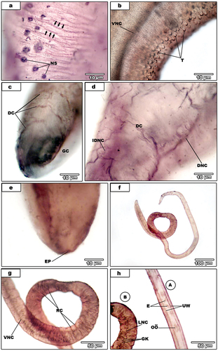
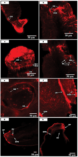
Figure 4. Confocal laser scanning micrographs of muscle activities in S. mansoni and S. haematobium.
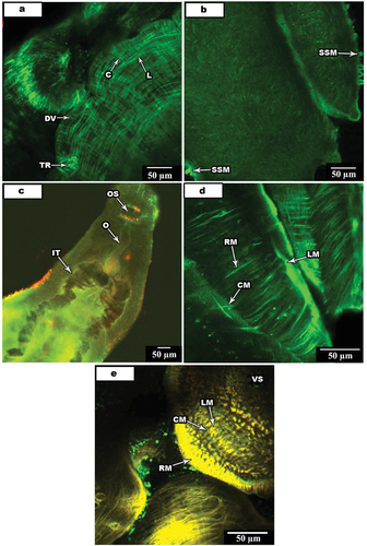
Figure 5. Confocal laser scanning micrographs of nerve activities in S. mansoni and S. haematobium.