Figures & data
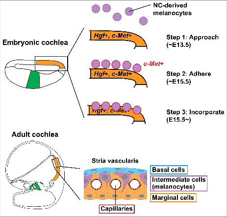
Figure 1. Schematic drawing of melanocyte incorporation into the stria vascularis of developing mouse cochlea. During mouse development, the prospective stria vascularis is specified in the embryonic cochlear epithelium (orange), distinct from the prospective organ of Corti (green). The stria vascularis consists of capillaries (crimson), basal cell layer (blue), intermediate cell (melanocyte) layer (purple) and marginal cell layer (orange). Neural crest (NC)-derived melanocytes approach the embryonic cochlear epithelium around embryonic day (E) 13.5 (Step 1). Around E15.5, the melanocytes adhere to the cochlear epithelium in the region of prospective stria vascularis (Step 2). These melanocytes start expressing c-Met (red). c-Met is weakly expressed in the cochlear epithelium and Hgf is expressed in the prospective stria vascularis (orange). The melanocytes then incorporate into the cochlear epithelium from basal turn of the cochlea (Step 3). The present study suggests that HGF-c-MET signaling in both melanocytes and cochlear epithelium is required for the initiation of the incorporation movement.