Figures & data
Figure 1 Diagram of the MDM2, MDMX, and p53. The structure of MDMX is similar to that of MDM2. Both MDMX and MDM2 contain four domains, including the amino-terminal hydrophobic p53 binding domain, the carboxy-terminal RING domain, the zinc finger domain and the central acidic domain. MDM2 has a unique NLS and NES signal sequence, which is related to the location of MDM2.
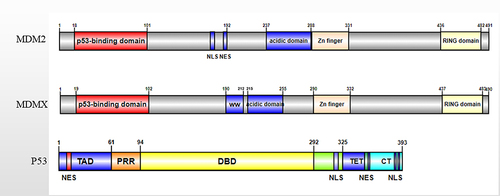
Figure 2 The schematic diagram of regulation of p53-related MDMX function. (A). N terminus p53-binding domain of MDMX can bind to p53 transactivation domain directly and inhibit p53 transactivation activity without promoting p53 degradation. (B). MDMX-MDM2 heterodimer is formed after the RING domains of them bind together, the heterodimer can promote MDM2-mediated p53 ubiquitination and degradation. (C). MDMX-MDM2 heterodimer can inhibit MDM2 ubiquitination, and increase MDM2 stability. MDM2 can promote MDMX ubiquitination and degradation too.
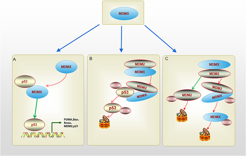
Table 1 Amplification and Overexpression of MDMX in Pancancer
Figure 3 Differential amplification and expression of MDMX in various cancers. (A). The expression levels of MDMX across pan-cancer. (B). CNV of MDMX across pan-cancer. (C). Correlation Analysis of MDMX CNV and Expression Levels (*P<0.05; **P<0.01; ***P<0.001; ns, P>0.05, not significant).
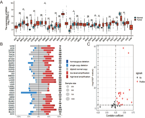
Table 2 A List of Current Available MDMX Inhibitors
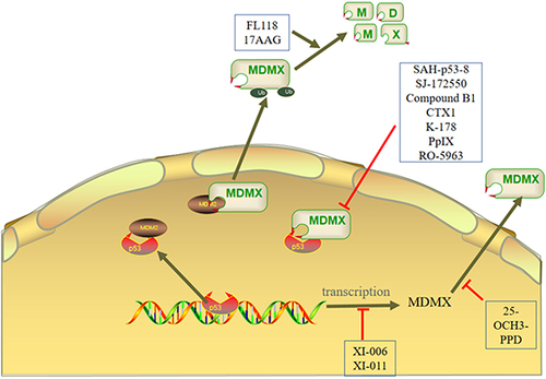
Figure 4 The mechanisms of MDMX inhibitors are mainly divided into three types. First, the inhibitor inhibits the expression of MDMX. Second, the inhibitor affects the formation of p53-MDM2/MDMX complex. Third, the inhibitor activates the E3 ligase activity of MDM2 to degrade MDMX.