Figures & data
Figure 1 Multiple subcutaneous lesions over different locations. (A and B) lesions in both elbows. (C) lesions in both knees. (D) lesions in buttocks. (E) lesions in Achilles tendons. (F) lesions over proximal interphalangeal joints.
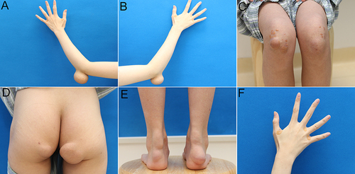
Figure 2 MRI imaging of xanthomas masses on left (A and B) and right (C and D) elbows. The masses revealed low signal intensity on T1-weighted images and low-high mixed signal intensity on T2-weighted images.
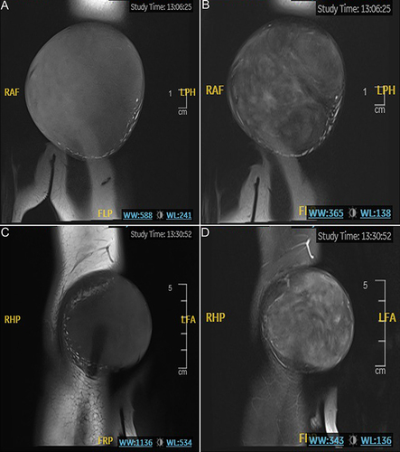
Figure 3 Intraoperative photograph of the lesions. (A and B) Resected xanthomas. (C) The sectional view of the xanthomas.
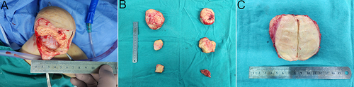
Figure 4 Pathological pictures of the lesion. (A) Hematoxylin/eosin analysis showed infiltration of foamy cells and scattered giant cells (x200). CD68-immunostaining (B), Ki67- immunostaining (C) and TFE3- immunostaining (D) of the xanthomas presented positive (x200).
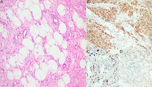
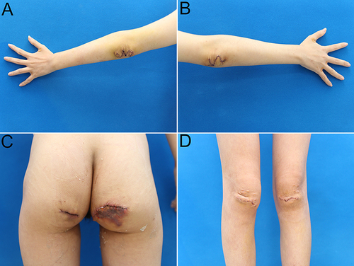
Figure 5 Status of the incisions two days after the operation. (A and B) elbows. (C) buttocks. (D) knees.