Figures & data
Figure 1 B3GALT5-AS1 was down-regulated in keloid tissues and KFs. (A) Clustered heatmap illustrating top 30 down-regulated lncRNAs in keloid tissues (n = 4) compared to normal skin tissues (n = 6). The red box indicates B3GALT5-AS1. (B) qRT-PCR validation of B3GALT5-AS1 expression in KFs and NFs. The data are expressed as the mean ± SD.
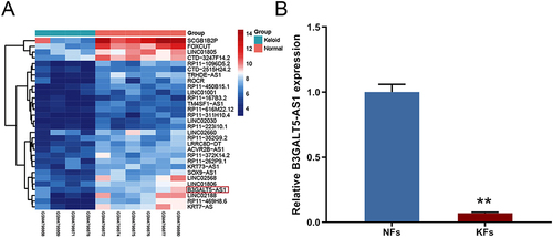
Figure 2 B3GALT5-AS1 suppressed KFs proliferation and metastasis in vitro. (A) qRT-PCR detected the B3GALT5-AS1 expression in KFs to demonstrate the infected efficacy of overexpressing or knocking down B3GALT5-AS1. (B) MTT assay was used to measure the cell viability of KFs. (C) A transwell assay was applied to determine cell invasion. The invasive cells were visualized using crystal violet staining. (D) Migration abilities were measured using wound healing assay. (E) Cell apoptosis was determined using flow cytometry. (F) Western blotting assays were subjected to detect the effect of B3GALT5-AS1 overexpression or knockdown on the tumor proliferative marker, apoptin, migration-associated proteins, and EMT marker expressions, full-length blots are presented in Supplementary Figure 1. The P-value was determined using Student’s t-tests. The data are expressed as the mean ± SD.
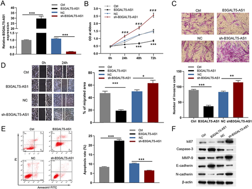
Figure 3 B3GALT5-AS1 inhibited the proliferation and invasion of KFs by repressing glycolysis. (A) The glucose consumption of KFs was examined in response to B3GALT5-AS1 overexpression or knockdown. (B) The lactate production in KFs was measured after B3GALT5-AS1 overexpression or knockdown. The ECAR (C) and OCR (D) in KFs were examined using Seahorse XF assay. (E) The glycolysis 2-DG attenuated weakened the effect of B3GALT5-AS1 on KFs proliferation. (F) The inhibition of B3GALT5-AS1 on cell invasion was restrained by 2-DG. The data are expressed as the mean ± SD. *p < 0.05, **p < 0.01 and ***p < 0.001. NS, no significant difference. KFs, primary keloid fibroblasts; B3GALT5-AS1, B3GALT5-AS1-overexpressed KFs; Ctrl, control of B3GALT5-AS1 overexpression; sh-B3GALT5-AS1, knockdown of B3GALT5-AS1 in KFs; NC, control of sh-B3GALT5-AS1.
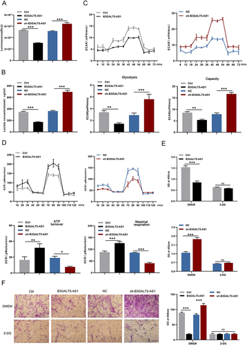
Figure 4 B3GALT5-AS1 interacted with HuR leading to its degradation. (A) The result of the lncLocator database revealed that B3GALT5-AS1 is localized predominantly in the cytoplasm. (B) The FISH assay identified the subcellular location of B3GALT5-AS1 in KFs. (C) qRT-PCR experiments demonstrated that B3GALT5-AS1 expression was primarily distributed in cytoplasm relative to the nucleus of KFs. (D) RNA pull-down and RIP assays verified the combination of B3GALT5-AS1 with HuR in KFs. (E) qRT-PCR and Western blotting determined the HuR mRNA and protein expression levels in KFs infected with B3GALT5-AS1 plasmid, si-B3GALT5-AS1, or their corresponding control. (F) Schematic structures of B3GALT5-AS1 and nine fragments. RIP experiments confirmed the interaction of F2 and HuR. (G) Design of B3GALT5-AS1 mutation. (H) RIP experiment of HUR with B3GALT5-AS1-wt or B3GALT5-AS1-mut. (I) Western blotting was applied to detect the effect of CHX treatment with or without MG 132 or chloroquine on attenuated HuR protein levels by B3GALT5-AS1 knockdown in KFs, full-length blots are presented in Supplementary Figure 1. (J) Ubiquitination experiments were adopted to determine the HuR degradation in KFs infected with sh-B3GALT5-AS1 or NC lentiviruses after treatment with MG 132. (K) Co-immunoprecipitation experiment was performed to evaluate the interaction between β-Trcp1 and HUR. ***p < 0.001. NS, no significant difference. KFs, primary keloid fibroblasts; B3GALT5-AS1, B3GALT5-AS1-overexpressed KFs; Ctrl, control of B3GALT5-AS1 overexpression; sh-B3GALT5-AS1, knockdown of B3GALT5-AS1 in KFs; NC, control of sh-B3GALT5-AS1.
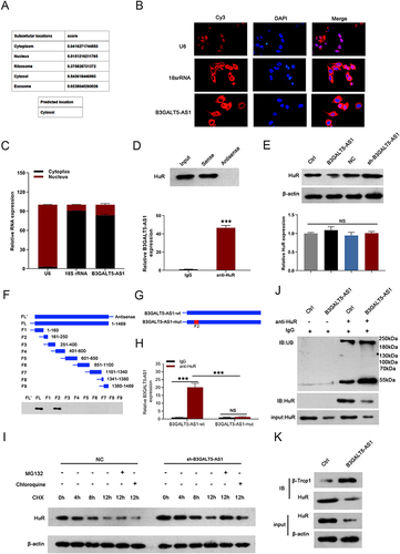
Figure 5 B3GALT5-AS1 degraded HuR to repress KFs proliferation and metastasis in vitro. (A) Western blotting detected the HuR protein expression in KFs after overexpressing HuR and B3GALT5-AS1. (B-D) MTT, Transwell, and wound healing assays illustrated that the inhibitory effects of B3GALT5-AS1 overexpression on the proliferation, invasion, and migration of KFs were significantly abolished by HuR overexpression. (E) HuR up-regulation suppressed cell apoptosis induced by B3GALT5-AS1 overexpression. (F) Western blotting analysis of Ki67, Caspase-3, MMP-9, E-cadherin, and N-cadherin protein expression, full-length blots are presented in Supplementary Figure 1. The data are expressed as the mean ± SD. **p < 0.01, and ***p < 0.001. KFs, primary keloid fibroblasts; B3GALT5-AS1, B3GALT5-AS1-overexpressed KFs; Ctrl, control of B3GALT5-AS1 or HuR overexpression; HuR, HuR-overexpressed KFs; HuR+ B3GALT5-AS1, KFs co-overexpressing HuR and B3GALT5-AS1.
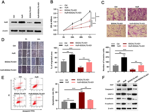
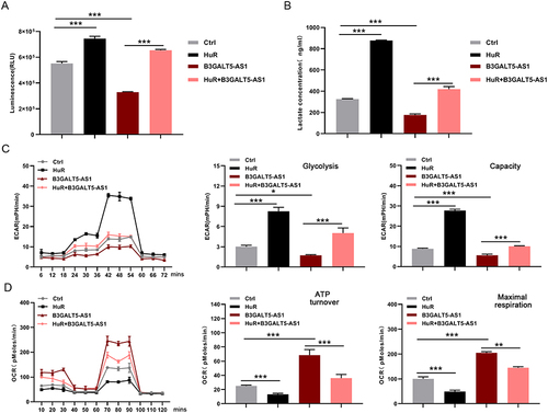
Figure 6 HuR attenuated the inhibitory effect of B3GALT5-AS1 on glycolysis in KFs. (A) The glucose consumption, lactate production (B), ECAR (C), and OCR (D) of KFs were detected after HuR, B3GALT5-AS1, or both HuR and B3GALT5-AS1 overexpression. The data are expressed as the mean ± SD. *p < 0.05, **p < 0.01, and ***p < 0.001. KFs, primary keloid fibroblasts; B3GALT5-AS1, B3GALT5-AS1-overexpressed KFs; Ctrl, control of B3GALT5-AS1 or HuR overexpression; HuR, HuR-overexpressed KFs; HuR+ B3GALT5-AS1, KFs co-overexpressing HuR and B3GALT5-AS1.