Figures & data
Figure 1 Clinical photos of the patients. (a–d) Diffuse infiltrated erythematous plaques observed on the dorsal side of the left forearm and back of the left hand. These plaques are accompanied by multiple insect-bite-like erosions, ulcers, papules, pustules, atrophic scars, and scabs within the affected area. (e and f) Following a 3-month treatment of oral terbinafine at a dosage of 250 mg/day combined with topical 1% naftifine-0.25% ketoconazole cream, an improvement in skin lesions was observed. Fibrotic scars emerged on the flexor aspect of the left arm as the skin lesions evolved.
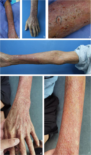
Figure 2 Histological Features of the Patient. (a) Pseudoepitheliomatous hyperplasia and granulomatous inflammation accompanied by localized necrosis and abundant inflammatory infiltration (H&E, magnification ×50). (b) Numerous morula-like formations highlighted through Periodic Acid-Schiff staining (magnification ×200). (c) Acid-fast stain: negative (magnification×50). (d) hexamine silver stains: positive (magnification ×200).
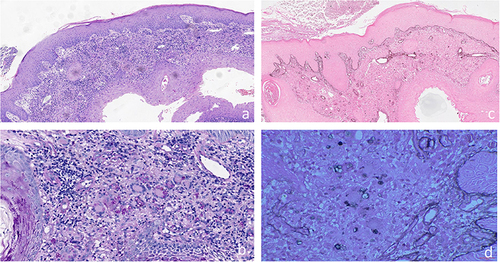
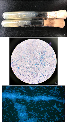
Figure 3 Colony Growth and Characteristics of Prototheca wickerhamii under Microscopy. (a) Smooth, creamy, white, and yeast-like colonies observed on SDA culture after 48 hours of incubation at 30°C. The characteristic endospore-containing sporangium of Prototheca wickerhamii. (b) Wet preparations stained with lactophenol cotton blue and (c) fluorochrome revealing the distinction from yeast due to the absence of budding.