
Figures & data
Figure 1 HE staining: A large number of nested Paget-like cells were found throughout the epidermis. The cytoplasm of tumor cells is rich, transparent and vacuolar. The basement membrane was intact, and a large number of lymph and plasma cells were infiltrated at the epidermal dermal junction. (A) Case1 (200X), (B) Case2 (200X), (C) Case3 (200X).
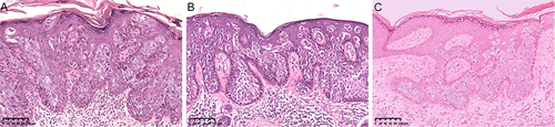
Figure 2 Immunohistochemical stain: We highlight the immunohistochemical staining of case 3. CK5/6 was positive for intact membrane, P63 was positive for nucleation, and CEA was negative. (A) CK5/6+ (200X), (B) P63+ (200X), (C) CEA- (200X).
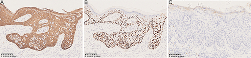
Table 1 Summary of Information and Immunohistochemical Stains of Patients with Pagetoid Squamous Cell Carcinoma