Figures & data
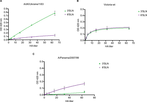
Figure 1 Influenza virus HA molecule from A/California/04/2009 (PDB: 3ube).
Notes: Each monomer in the (A) trimer is indicated in color; the front view shows the HA1 (green or gray) and HA2 subunits (red or orange). A closer view of the (B) globular head and the interaction with ligand LSTc; in purple are shown some of the amino acid residues at the RBS that directly interact with the sialic acid-like receptor. Images were generated with Yasara software version 17.1.28.
Abbreviations: HA, hemagglutinin; LSTc, sialylneolacto-N-tetraose c; RBS, receptor-binding site; PDB, Protein Data Bank.
Abbreviations: HA, hemagglutinin; LSTc, sialylneolacto-N-tetraose c; RBS, receptor-binding site; PDB, Protein Data Bank.
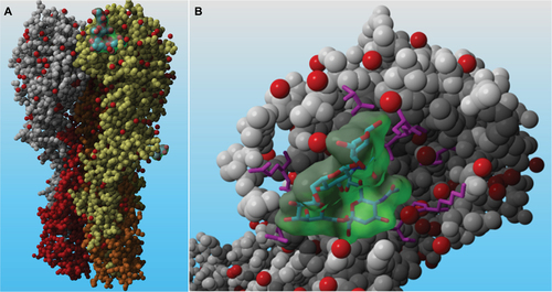
Figure 2 Schematic representation of the methods and techniques used throughout influenza virus history to identify the receptor type recognition.
Table 1 Residues at the RBS of the HA protein of H1N1 viruses isolated from humans, avian, or swine
Figure 3 Binding of influenza H3 viruses representing different stages of the subtype evolution prior to and since its emergence in humans.
Notes: Viruses were analyzed by cross-link binding assay using receptor mimics 3′SLN and 6′SLN. Virus binding to polymers was detected with virus subtype-specific antiserum.Citation64 (A) Binding of an AIV (H3N8) to synthetic polymers. The virus shows preference for 3′SLN. (B) Binding of early human IAV (H3N2) to synthetic polymers. The virus shows dual preference for 3′SLN and 6′SLN. (C) More recent human H3N2 viruses switch the receptor preference for 6′SLN and reduced affinity for the avian-like polymers 3′SLN.
Abbreviations: 3′SLN, 3′-sialyl-N-acetyllactosamine trisaccharide; 6′SLN, 6′-sialyl-N-acetyllactosamine; HA, hemagglutinin.
Abbreviations: 3′SLN, 3′-sialyl-N-acetyllactosamine trisaccharide; 6′SLN, 6′-sialyl-N-acetyllactosamine; HA, hemagglutinin.