Figures & data
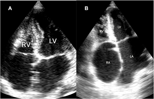
Figure 1 AP4 view. (A) Shows the main feature of EMF: apical and left ventricular outflow tract fibrosis, huge left atrium and left ventricular cavity obliteration. (B) Shows the mitral valve regurgitation (MR).
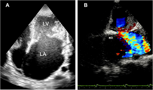
Figure 2 LAP view showing huge left atrium (LA), endomyocardial fibrous shelf (EFS), anterior mitral valve leaflet (AMVL), pericardial effusion (PE), and left ventricle (LV).
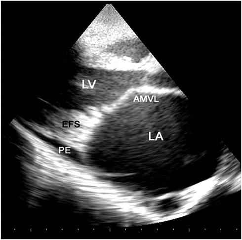
Figure 3 M-Mode view showing the layering of the posterior LV wall with a cleavage line between the endocardium (E) and the myocardium (M).
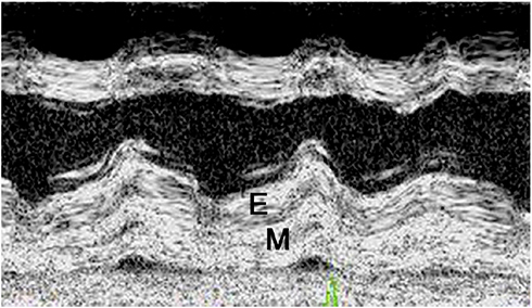
Figure 4 Short axis view showing endocardial fibrosis (E) engulfing the posterior papillary muscle. Calcification of the pericardium and pericardial effusion (P) are seen.
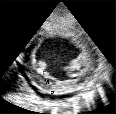
Figure 5 AP4 view: (A) view of a case of right ventricular (RV) EMF. (B) Shows a case of biventricular EMF of both RV and LV.