
Figures & data
Figure 1 Surgical layout for the implementation of LVATS. (A) Configuration for left lung surgery. (B) Arrangement for right lung surgery, with appropriate modifications for operative access based on the surgeon’s preference. (C) Essential instruments used in LVATS.
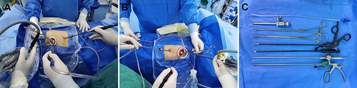
Figure 2 Appropriate instruments inserted through a 5mm incision enhance surgical exposure, enable efficient tissue mobilization, and allow for the precise deployment of a surgical stapler. (A) Transecting the oblique fissure. (B) Circling the bronchus of the right upper lobe. (C) Transecting the bronchus of the right upper lobe. (D) Circling the arterial branch of the right upper lobe. (E) Transecting the arterial branch of the right upper lobe. (F) Circling the vein of the right upper lobe. (G) Transecting the vein of the right upper lobe. (H) Lymphadenectomy of the 2nd and 4th nodal stations. (I) Lymphadenectomy of the 7th nodal station.
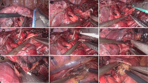
Figure 3 Location and details of the incisions. (A) A 2.5 cm utility incision was made in the 4th intercostal space, and a 5 mm trocar was placed in the 7th intercostal space. (B) A 16 Fr drainage tube was inserted into the 7th intercostal space.
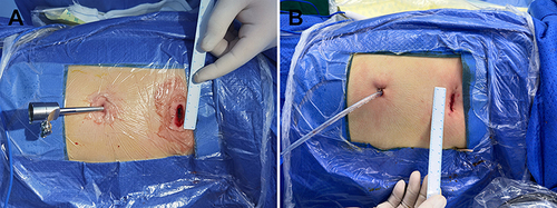
Figure 4 Coaxial manipulation of instruments during UVATS. (A) Dissection at the pulmonary hilum for clear access. (B) Lymphadenectomy of the 10th nodal station. (C) Lymphadenectomy of the 7th nodal station. (D) Mobilization of the right upper lobe’s arterial branch to secure vascular control. (E) Mobilization of the right upper lobe bronchus for bronchial management. (F) Mobilization of the right upper lobe vein for venous control.
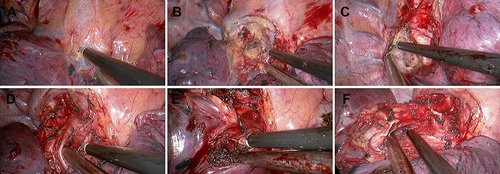
Table 1 Clinical Features Before and After Propensity-Matched
Table 2 Comparison on Perioperative Outcomes